|
[Home]
[Up]
[Parenchyma Images]
[Ragweed pith]
[Primary pit fields]
[Primary pit fields]
[Wall, face view]
[Nucleate cells]
[Compact parenchyma]
[Sclerenchyma]
[Intercellular spaces]
[Leaf, xs]
[Chlorenchyma]
[Shoot tip, ls]
[Apical meristem]
[Vascular cambium]
[Secretory duct]
[Resin canal]
[Clusia duct]
[Root cortex]
[Leaf aerenchyma]
[Air chambers]
[Air chambers]
[Aerenchyma, Acorus]
[Stellate cells]
[Epidermis]
[Endodermis]
[Phloem, ls]
[Phloem, xs]
[Transfer cells]
[Bean cotyledon]
[Acorn, starch]
[Potato starch]
[Ice plant cell]
[Tannin cell]
[Small cells]
| |
Chapter 3. Parenchyma.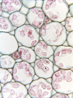
The figures presented here were
selected to illustrate aspects of parenchyma cells and tissues, and the legends
were written to complement the more complete discussion of parenchyma presented
in Chapter 3 (pages 43 to 51) in the textbook Plant Anatomy by J. D. Mauseth,
published by Cummings & Hathaway.
Click here for a set of links to the micrographs of this
chapter.
Potato parenchyma with starch grains
|