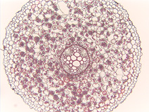
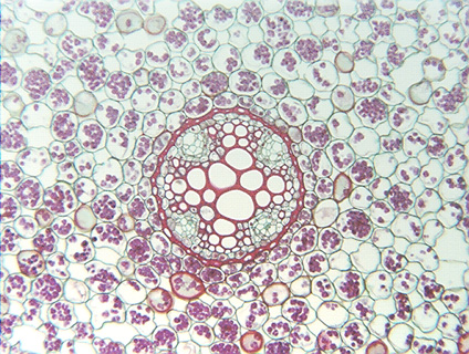
Fig. 3.3-1a and b. Transverse section of root of buttercup (Ranunculus). The circular structure in the center of the large micrograph contains the conducting tissues of the root, but now we are concerned only with the outer tissue, the root cortex. All these cortex cells are parenchyma, and because the bulk of the root consists of this (see the inset), this is structural parenchyma. It is also storage parenchyma because most cells are full of starch grains (stained purple). The starch grains (amyloplasts) developed from proplastids and are so abundant that nuclei are hidden. Some cells have few or no starch grains, probably because some grains fell out while the slide was being stained, after the cells had been cut open during microtoming. Because the starch grains are so abundant, it is easy to tell which areas are cells and which are intercellular spaces, and this tissue has a large percentage of its volume as intercellular space. Many botanists would consider the intercellular spaces here sufficiently abundant to call this aerenchyma, others would consider it a bit too compact -- there is no universal definition of aerenchyma.