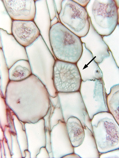
 Fig. 3.1-4. Transverse
section of Aristolochia stem. The parenchyma cell in the very center
(arrow) appears to be filled with a weblike mesh, but in fact we are looking at either
the front or the back wall, and virtually the entire wall is a set of primary
pit fields. The whitish areas that appear to be holes are just areas
where the primary wall is particularly thin and filled with plasmodesmata (we
cannot see plasmodesmata with ordinary brightfield light microscopy, but these
thin areas typically contain high concentrations of plasmodesmata). The darker
material that forms the mesh is just primary wall of ordinary thickness. If the
microscope illuminator were turned bright enough, it too would appear whitish.
Fig. 3.1-4. Transverse
section of Aristolochia stem. The parenchyma cell in the very center
(arrow) appears to be filled with a weblike mesh, but in fact we are looking at either
the front or the back wall, and virtually the entire wall is a set of primary
pit fields. The whitish areas that appear to be holes are just areas
where the primary wall is particularly thin and filled with plasmodesmata (we
cannot see plasmodesmata with ordinary brightfield light microscopy, but these
thin areas typically contain high concentrations of plasmodesmata). The darker
material that forms the mesh is just primary wall of ordinary thickness. If the
microscope illuminator were turned bright enough, it too would appear whitish.