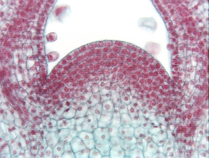
 Fig.
3.2-4.
Longitudinal section of the shoot apical meristem
of Coleus. This is a magnification of the apical meristem shown in Fig.
3.2-3. All cells here are parenchyma cells involved in the synthesis of new
cells. Each cell is almost filled by a prominent round, red-stained nucleus (in
some nuclei you can see a dark red, dot-like nucleolus). These cells, like most
apical meristem cells, are small, not much larger than the nucleus. All
organelles are present, but too small to be seen: plastids are present as small
proplastids not large chloroplasts, vacuoles are small and scattered rather than
being coalesced into a large central vacuole, and all other organelles are never
visible by ordinary light microscopy. Because these meristematic cells are so
small, cell division -- cytokinesis -- can occur quickly because the
phragmoplast and cell plate do not have to grow to a large size before they meet
the side walls.
Fig.
3.2-4.
Longitudinal section of the shoot apical meristem
of Coleus. This is a magnification of the apical meristem shown in Fig.
3.2-3. All cells here are parenchyma cells involved in the synthesis of new
cells. Each cell is almost filled by a prominent round, red-stained nucleus (in
some nuclei you can see a dark red, dot-like nucleolus). These cells, like most
apical meristem cells, are small, not much larger than the nucleus. All
organelles are present, but too small to be seen: plastids are present as small
proplastids not large chloroplasts, vacuoles are small and scattered rather than
being coalesced into a large central vacuole, and all other organelles are never
visible by ordinary light microscopy. Because these meristematic cells are so
small, cell division -- cytokinesis -- can occur quickly because the
phragmoplast and cell plate do not have to grow to a large size before they meet
the side walls.