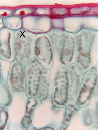
 Fig. 3.2-2.
Transverse section of leaf of magnolia (Magnolia). Palisade parenchyma at
a higher magnification than that of Fig. 3.2-1. Thin primary walls are easy to
see as fine blue lines; the protoplasts shrank somewhat when the samples were
treated with alcohol, so there is a bit of empty, white space between the wall
and protoplast in some cells. This artifact makes it easier to see the wall.
Several nuclei are present, one is round, others are oval or flattened; all have
been stained gray and each has one or two small, dark red nucleoli. Chloroplasts
form a single uniform layer, packed together so closely that they cover almost
all the wall. In the cell marked X,
the front wall is present in the section (we can tell it is the front wall, not
the back wall, because the chloroplasts that lie against it are partially
obscuring the nucleus; if it were the back wall, the nucleus would hide the
chloroplasts). In this cell, you can see that chloroplasts are round in face
view. In other cells, side walls come directly up at us, and chloroplasts are
visible in side view; in most areas, it is easy to see each one individually. In
the areas where they are not so distinct, it is because they are crowded
together.
Fig. 3.2-2.
Transverse section of leaf of magnolia (Magnolia). Palisade parenchyma at
a higher magnification than that of Fig. 3.2-1. Thin primary walls are easy to
see as fine blue lines; the protoplasts shrank somewhat when the samples were
treated with alcohol, so there is a bit of empty, white space between the wall
and protoplast in some cells. This artifact makes it easier to see the wall.
Several nuclei are present, one is round, others are oval or flattened; all have
been stained gray and each has one or two small, dark red nucleoli. Chloroplasts
form a single uniform layer, packed together so closely that they cover almost
all the wall. In the cell marked X,
the front wall is present in the section (we can tell it is the front wall, not
the back wall, because the chloroplasts that lie against it are partially
obscuring the nucleus; if it were the back wall, the nucleus would hide the
chloroplasts). In this cell, you can see that chloroplasts are round in face
view. In other cells, side walls come directly up at us, and chloroplasts are
visible in side view; in most areas, it is easy to see each one individually. In
the areas where they are not so distinct, it is because they are crowded
together.
Organelles such as mitochondria, endoplasmic reticulum and dictyosomes are also present, but are never visible by ordinary light microscopy.
Notice that the epidermal cells have thick walls stained a very dark red. Most of that is due to the water-proofing chemical cutin in the walls, but the main point to notice now is that the primary walls of palisade parenchyma cells are extremely thin compared to those of epidermis cells. The layer of rather empty-looking cells between epidermis and palisade parenchyma is called hypodermis.