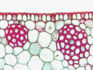
 Fig. 3.1-7. Transverse
section of leaf of dracaena (Dracaena). This section contains both sclerenchyma
(the two masses of cells with red-stained walls, but not the epidermis along the
top) and parenchyma (all the other cells).
This figure shows the difference between the thin primary walls of parenchyma
cells and the thick secondary walls of the fibers. The primary walls of the
parenchyma cells do not contain lignin, so they have not taken up the red stain,
but both the primary and the secondary walls of the fiber cells are lignified
and have been stained so intensely that the primary walls of the fibers cannot
be distinguished from the secondary walls.
Fig. 3.1-7. Transverse
section of leaf of dracaena (Dracaena). This section contains both sclerenchyma
(the two masses of cells with red-stained walls, but not the epidermis along the
top) and parenchyma (all the other cells).
This figure shows the difference between the thin primary walls of parenchyma
cells and the thick secondary walls of the fibers. The primary walls of the
parenchyma cells do not contain lignin, so they have not taken up the red stain,
but both the primary and the secondary walls of the fiber cells are lignified
and have been stained so intensely that the primary walls of the fibers cannot
be distinguished from the secondary walls.