|
About
the lab:
The Szaniszlo Laboratory is a research
laboratory located on the first floor of the Experimental Science Building
(ESB 109) at the University of Texas at Austin. The lab is part of the Section of
Molecular Genetics and Microbiology and is headed by Dr. Paul J. Szaniszlo. The
personnel in the lab consist of Dr. Szaniszlo, 2 post-docs, 4 graduate students, as
well as some lab assistants. Click on "People"
to learn more about them.
About our
research:
The Szaniszlo Lab's research is
primarily in the elucidation of the mechanisms of fungal cell development, specifically
that of the fungus, Wangiella dermatitidis (Wd). Wangiella is used
as a model to identify cell-wall virulence factors among dematiaceous (melanized) fungal
pathogen of humans. Specific emphasis is placed on the cell-wall because it is a
boundary between host and pathogen, an ultimate determinant of morphology, known to
represent a virulence factor, and is a potential target for the design of antifungal
drugs. Particular focus is on the systems leading to chitin localization and melanin
enrichments of cell walls in the polarized and nonpolarized vegetative phenotypes known to
be expressed by this polymorphic fungus under conditions of human infection.
Thus, the lab's current research is designed to provide
insights about how the four chitin synthase (WdCHS) structural genes of Wangiella
contribute to its pathogenicity and virulence. The specific aims of the laboratory are
- To study the WdCHS genes, establish how their expression is
regulated, and identify factors that control the time, place and function of their
products, with a focus on the mechanisms involved in the very high expression of WdCHS3
at 37�C and under other conditions that influence growth, development and differentiation
during infections;
- To establish the individual and collective importance of the four
WdChs isozymes and their chitin products to yeast, isotropic and hyphal growth, and their
contributions, if any, to pathogenicity and virulence as augmented by melanin and periods
of isotropic enlargement, and with a focus on the potential for WdChs3p to be a
particularly vulnerable antifungal target
- To continue to extend the concepts revealed with this model
phaeohyphomycotic fungus to a limited number of other dematiaceous pathogens of humans
What
is Wangiella dermatitidis?
Wangiella
dermatitidis, also often called Exophiala dermatitidis, is a
dematiaceous fungus known for causing a variety of infections known collectively as
"phaeohyphomycosis". It is a monotypic species in the form-phylum Fungi
Imperfecti (Deuteromycetes). What distinguishes it as a dematiaceous fungus is the
black pigment seen in the cell walls of all its yeast cells, hyphae, multicellular forms,
and spores. Wangiella is also a polymorphic organism, taking on a variety of cell
types, which include the typical budding yeast cell, isodiametric cell, moniliforme
hyphae, true hyphae, and conidia. It grows as a yeast in rich liquid medium, but has
been shown to convert to other forms by modifying growth conditions.
Wangiella is
readily found in the environment worldwide, particularly in soil. Although
ubiquitous, it does not seem to be a major cause of mycoses (fungal disease).
What
is phaeohyphomycosis?
| Phaeohyphomycosis
is a term that was created in 1974 to include several infections that are
caused by many but not all mycotic diseases caused by black fungi.
Phaeohyphomycosis can be superficial (on the surface of the skin, hair, or nails),
cutaneous (involving the skin immediately below the outer epidermis), subcutaneous
(involving fatty tissue, connective tissue, or muscle tissue), or systemic (involving the
circulatory and/or lymphatic system). The more serious life-threatening, systemic
phaeohyphomycoses, mostly occurs with immunosuppressed individuals. Although
phaeohyphomycosis traditionally has been most associated with dermotrophic forms of
disease, emerging systemic forms are being detected in increasing number. For
example, predisposing factors for systemic infections with W. dermatitidis
include cystic fibrosis, lymphocytic leukemia, diabetes mellitus, bronchiectasis,
rheumatoid arthritis and catherization. For
more information on the clinical aspect of this topic, see Matsumoto, T., Matsuda, T.,
McGinnis, M.R., and Ajello, L. 1992. Clinical and mycological spectra of Wangiella
dermatitidis infections. Mycoses 36: 145-155. |
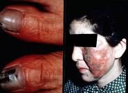 |
| Ungual
phaeohyphomycosis (L), Cutaneous phaeohyphomycosis (R). courtesy of Dr. Tadahiko Matsumoto, Dept. of Dermatology, Toshiba Hospital,
6-3-22 Higashi-oi, Shinagawa-ku, Tokyo 140, Japan |
Help!
I'm not a scientist! Can you translate any of this for me?
Wangiella dermatitidis, the subject of this lab's research
is a black (melanized) fungus. Fungi (the plural of fungus) are a kingdom of
organisms that include your garden variety mushroom (Agaricus brunnescens),
baker's yeast (Saccharomyces cerevisiae), the causative agents of athlete's
foot (E. floccosum, T. mentagrophytes, and T. rubrum), the cause of
yeast infections (Candida albicans), the mold you might see
growing on your bread or cheese (Rhizopus stolnifer), or the mold (Penicillium
notatum) that produces the medicine used to combat an infection. Most people
are unaware of all the roles(both positive and negative) that fungi play in our
lives.
Wangiella has been known to cause
disease that can exhibit a wide variety of symptoms. Although it can certainly be
life-threatening, that in itself is not the only reason for researching this organism.
Fungi have many common attributes with "higher" life forms. Many
biochemical and molecular properties of cell growth and regulation are similar.
Thus, fungi like Wangiella can be used as models to investigate fungi and
other life forms that are not as easily studied at biochemical and molecular levels.
Wangiella has been demonstrated to be a
paradigm for mycotic infections caused by black fungi. This means that it alone
elicits all the broad varieties of disease manifestations in humans which are
characteristic of those presented by any other melanized fungus.. Understanding the
mechanisms that control the growth of these fungi will provide insights into their
pathogenicity and virulence and may lead to improved treatments.
How
do I get around this site?
The Szaniszlo
Lab website is navigable by clicking on one of the text selections on the black region to
the left of the page. You will notice that the selections will highlight when you
move the cursor over them. With your mouse pointer over the selection, click on the
text to jump to that page. The choices are Introduction,
People, Publications, Protocols, Images, Courses,
Links, and Search. If your browser
is having trouble with the selections on the left, you can still make your selections
using the same text selections at the bottom of every page. You can return to the
Szaniszlo Lab's main page at any time by clicking on "the
Szaniszlo Lab" at the top right hand corner of any page.

How
do I contact the Szaniszlo Lab
You can
contact the Szaniszlo Lab in three ways. You can send us a snail-mail, an e-mail, or
call the lab:
|
Szaniszlo Lab
Section of Molecular Genetics and Microbiology ESB 109
Campus Mail Code: A5000
University of Texas at Austin
Austin, Texas 78712-1095E-mail:
Dr. Paul J. Szaniszlo: pjszaniszlo@mail.utexas.edu
Website administrator: venture@mail.utexas.edu
Laboratory phone number:
(512) 471-7080
For the e-mail addresses of the rest of the lab
group, please check the People section. |
|