|
[Home]
[Up]
[Dicot stem]
[Monocot stem]
[Broad pith]
[Weak stem]
[Monocot fiber sheaths]
[Ordinary cortex]
[Aerenchyma hypodermis]
[Aerenchyma cortex]
[Aerenchyma cortex 2]
[Stem endodermis]
[Palisade cortex]
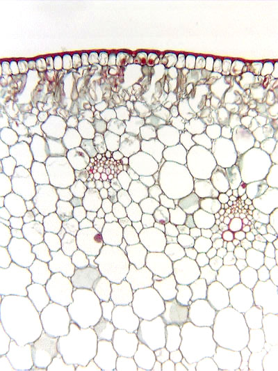
[Cortical bundle]
[Capped cortical bundles]
[Collapsible cortex]
[Perimedullary fibers]
[Conjunctive tissue, paren.]
[Torn pith]
[Hollow pith]
[Medullary bundles]
[Typical dicot bundle]
[Vascular ring]
[Typical monocot bundle]
[Amphivasal bundle]
[Corn vascular bundle]
[Clintonia bundles]
[Protoxylem]
[Metaxylem]
[Metaxylem parenchyma]
[Metaxylem fibers]
[Internal phloem]
[Internal phloem, mag]
[Developing metaxylem]
[Primary phloem]
[Phloem fiber cap]
[Developing fibers]
| |
 Chapter
11. Stems. Chapter
11. Stems.
The figures presented here were selected to illustrate aspects of stem
anatomy, and the legends were written to complement the
more complete discussion presented in Chapter 11 (pages 201 to 230)
in the textbook Plant Anatomy by J. D. Mauseth, published by Cummings
& Hathaway.
Click here for a set of links to the micrographs of this
chapter.
Transverse
section of stem of castor bean (Ricinus)
|