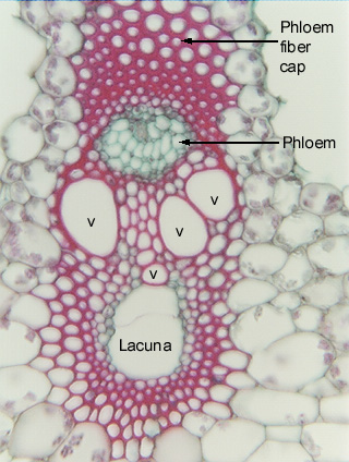
 Fig.
11.5-10. Transverse section of sedge stem (Scirpus americana).
Unlike the primary xylem of the previous two micrographs, this
primary xylem has almost no parenchyma. Instead, almost all the xylem cells that
are not vessels are fibers. The conjunctive tissue consists of
parenchyma cells, and much of the mechanical strength that supports the stems is
provided by the fibers of these bundles, along with a fibrous hypodermis (not
shown here).
Fig.
11.5-10. Transverse section of sedge stem (Scirpus americana).
Unlike the primary xylem of the previous two micrographs, this
primary xylem has almost no parenchyma. Instead, almost all the xylem cells that
are not vessels are fibers. The conjunctive tissue consists of
parenchyma cells, and much of the mechanical strength that supports the stems is
provided by the fibers of these bundles, along with a fibrous hypodermis (not
shown here).
The lacuna was formed by the tearing of protoxylem tracheary elements. Notice that, like the vascular bundles of many dicots, the phloem is accompanied by a thick cap of primary phloem fibers, which here makes up part of the bundle sheath. By being located to the exterior of the phloem, they provide some protection against insects with sucking mouth parts.
The two large open spaces on either side of the phloem (the labels are in the one on the right) are large intercellular spaces that run through the cortex, parallel to the surface of the stem.