|

(to view an enlarged
version of the image, click on the image. A high-res version should appear, if
available. Load times may take a while, depending on your connection speed.
Click your browser's "back" button to return to this gallery.)
The subject of study
at the Szaniszlo Lab:
Wangiella
dermatitidis
 |
|
 |
|
 |
|
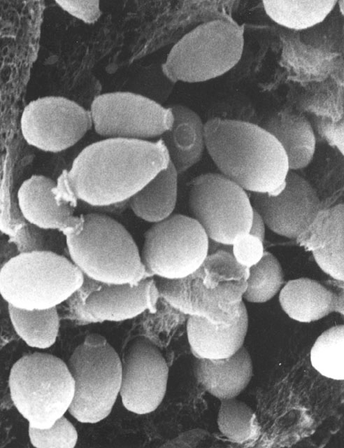
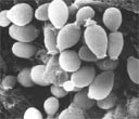
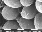
Wangiella,
wt,
25°C, budding yeast with scars (McIntosh) |
|
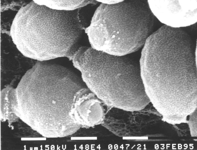
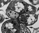
Wangiella,
wt,
25°C, budding yeast with scars, close up (McIntosh) |
|
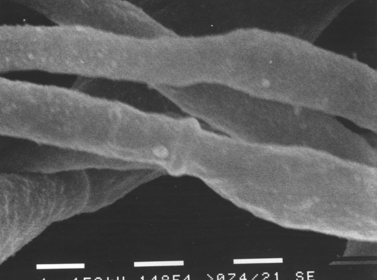
Wangiella,
wt,
25°C, budding yeast with scars, close up, different view (McIntosh) |
 |
|
 |
|
 |
|
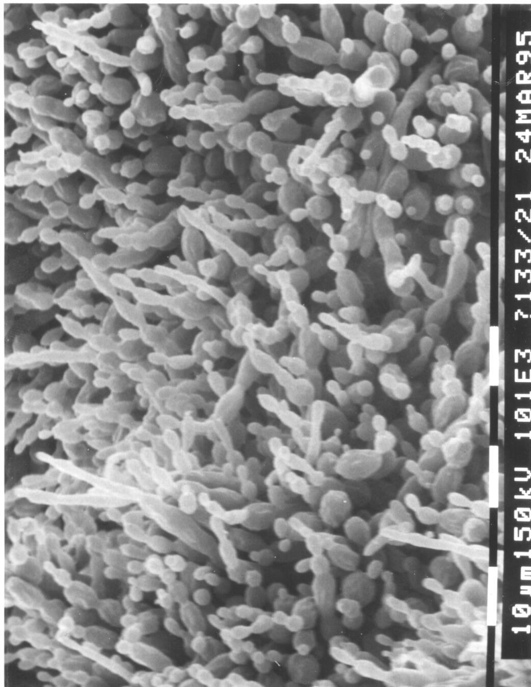
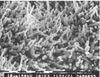
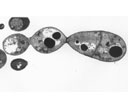
Wangiella,
Hf1,
28°C, length of hyphae (McIntosh) |
|
Wangiella, wt,
37°C, budding (McIntosh) |
|
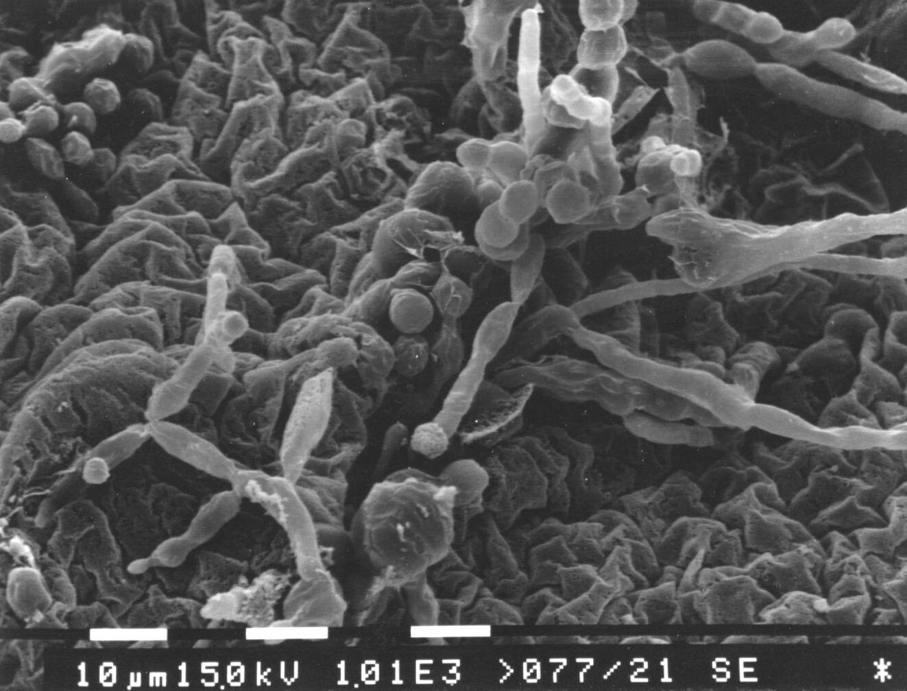
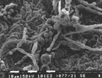
Wangiella,
Hf1,
isotropic cells among hyphae (McIntosh) |
 |
|
 |
|
 |
|
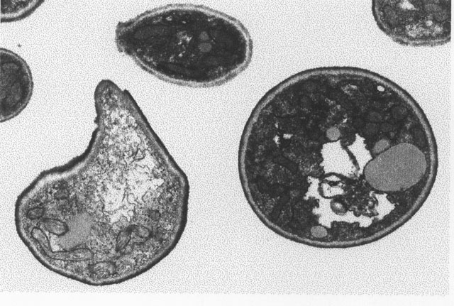
Wangiella, wdchs3
mutant, 37°C |
|
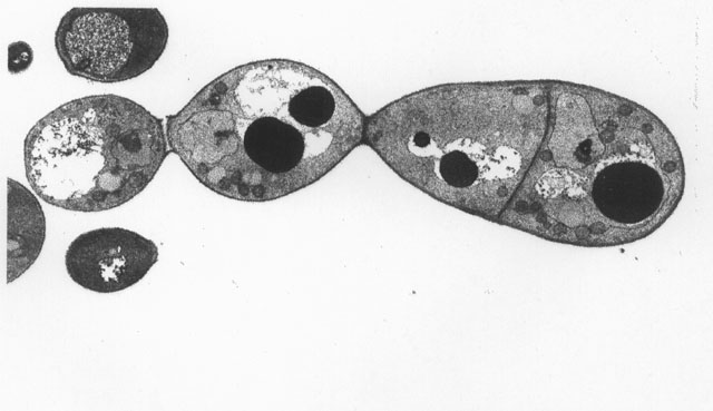
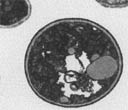
Wangiella,
multicellular, sclerotic form. |
|
Wangiella, Hf1,
37şC |
|