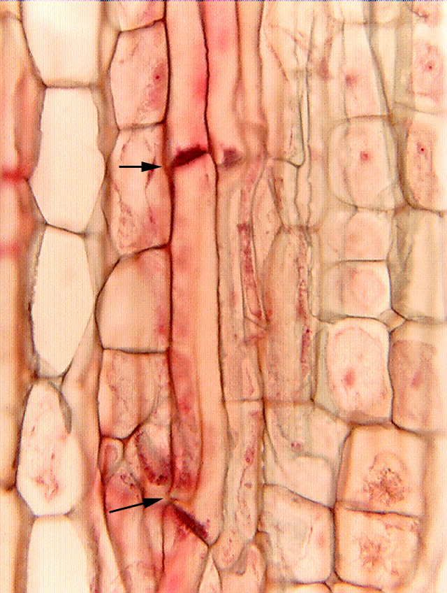
 Fig.
8.2-1. Longitudinal section of stem of milkweed (Asclepias).
The two arrows indicate the sieve plates at the ends of a sieve tube member; the
two adjacent members of this same tube are located above the upper sieve plate
and below the lower one. Notice especially that the lower
sieve plate is tilted and that the sieve tube members are a little bulbous
there, a bit bent. It looks as if the sieve tube members – after
they became mature – elongated a tiny bit, just enough that their ends had to
bend out of alignment. This is typical of phloem, and is one
of your best criteria for identifying sieve tube members in
longitudinal section. In transverse section, sieve tube members appear empty,
but this does not usually help in longitudinal sections because the tubes are
slender relative to the thickness of the section. There is almost always a thin
layer of other cells either in front of or behind the sieve tube member we are
trying to examine. It is necessary to focus a microscope up and down while
watching carefully to be sure that the sieve tube member is basically empty
(other than the thin layer of cytoplasm along the cell wall). Rather than doing
that, finding the tilted sieve plates is much faster, easier and more reliable.
Fig.
8.2-1. Longitudinal section of stem of milkweed (Asclepias).
The two arrows indicate the sieve plates at the ends of a sieve tube member; the
two adjacent members of this same tube are located above the upper sieve plate
and below the lower one. Notice especially that the lower
sieve plate is tilted and that the sieve tube members are a little bulbous
there, a bit bent. It looks as if the sieve tube members – after
they became mature – elongated a tiny bit, just enough that their ends had to
bend out of alignment. This is typical of phloem, and is one
of your best criteria for identifying sieve tube members in
longitudinal section. In transverse section, sieve tube members appear empty,
but this does not usually help in longitudinal sections because the tubes are
slender relative to the thickness of the section. There is almost always a thin
layer of other cells either in front of or behind the sieve tube member we are
trying to examine. It is necessary to focus a microscope up and down while
watching carefully to be sure that the sieve tube member is basically empty
(other than the thin layer of cytoplasm along the cell wall). Rather than doing
that, finding the tilted sieve plates is much faster, easier and more reliable.
In this side view, we cannot actually see the sieve pores. The fixation here was so gentle that no P-protein plugs formed; if they had, they would also help in identifying these sieve tube members.
Companion cells? Trying to identify those in longitudinal section is very difficult, and usually requires sections that are so thin our view is not obscured by cells in front of or behind the ones we are examining.