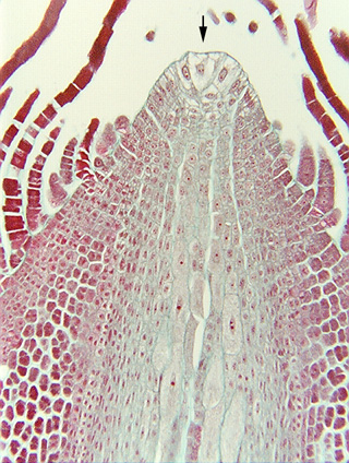
 Fig. 6.3-1a and b.
Longitudinal section of a shoot tip of a fern (Nephrolepis). Ferns have a
shoot apical meristem that contains a prominent single apical cell (arrow),
which is visible here as the uppermost, central cell at the tip of the shoot.
Notice that there are two cells on either side of the apical cell, and another
cell below it (mostly distinguishable because their nuclei are visible). The
three cells plus the apical cell altogether make up a pyramidal complex that has
the outline of an apical cell, and that is because the surrounding cells were
recently parts of the apical cell, and became cells during the last few
divisions of the apical cell. Below the apex is a column of lightly-stained
cells that will develop into pith, surrounded by dark red cells that will become
cortex. The rows of cells projecting upward are trichomes.
Fig. 6.3-1a and b.
Longitudinal section of a shoot tip of a fern (Nephrolepis). Ferns have a
shoot apical meristem that contains a prominent single apical cell (arrow),
which is visible here as the uppermost, central cell at the tip of the shoot.
Notice that there are two cells on either side of the apical cell, and another
cell below it (mostly distinguishable because their nuclei are visible). The
three cells plus the apical cell altogether make up a pyramidal complex that has
the outline of an apical cell, and that is because the surrounding cells were
recently parts of the apical cell, and became cells during the last few
divisions of the apical cell. Below the apex is a column of lightly-stained
cells that will develop into pith, surrounded by dark red cells that will become
cortex. The rows of cells projecting upward are trichomes.
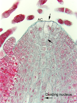 Figure b is a high magnification view of the apex of Nephrolepis.
The apical cell (AC) is the triangular cell. It seems somewhat small, perhaps
because it has just finished a cell division or perhaps because the section is
passing through one of the narrow parts of its pyramidal
shape. To its right is
a large square cell (arrow) with a prominent nucleus, and below that cell is an
inconspicuous cell about the same size (arrow). Those two cells are sister
cells, produced by the transverse division of a long narrow cell that had been
cut off from the side of the apical cell. Soon, the apical cell will cut off
another long narrow cell along its left side, and then that cell will divide
into two progeny cells by a transverse wall, creating a stack of two cells
similar to the stack that already occurs on the right side.
Figure b is a high magnification view of the apex of Nephrolepis.
The apical cell (AC) is the triangular cell. It seems somewhat small, perhaps
because it has just finished a cell division or perhaps because the section is
passing through one of the narrow parts of its pyramidal
shape. To its right is
a large square cell (arrow) with a prominent nucleus, and below that cell is an
inconspicuous cell about the same size (arrow). Those two cells are sister
cells, produced by the transverse division of a long narrow cell that had been
cut off from the side of the apical cell. Soon, the apical cell will cut off
another long narrow cell along its left side, and then that cell will divide
into two progeny cells by a transverse wall, creating a stack of two cells
similar to the stack that already occurs on the right side.
Note the dividing nucleus near the bottom of the micrograph.