
Images Gallery
Click on an image to view a larger high-resolution image, then scroll to view details. Click outside the high-resolution image to close it.
- Retina
- Macular Pigment
- Electrode Track Marking
Galleries:
Retina
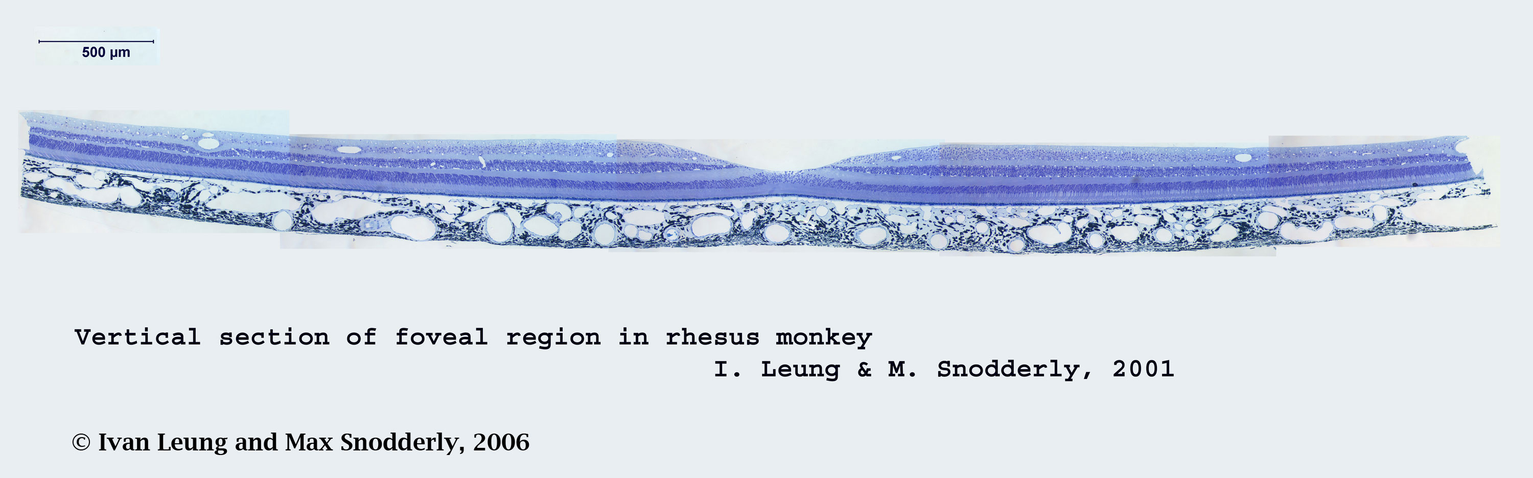
Arrangement of the cellular layers in the foveal region of a diurnal primate retina. Composite of 5 micrographs of a vertical section through the fovea of a rhesus monkey (12176) taken at 200X magnification. The tissue was perfused with 4% paraformaldehyde, 0.5 % glutaraldehyde and 4% polyvinyl-pyrrolidone (PVP) prior to embedment in methacrylate. The two-micron-thick section was stained with toluidine blue to visualize the cell nuclei. Other details of tissue preparation are available in Leung IY-F, et al. Invest Ophthalmol Vis Sci. 2004; 45: 3244-3256. Note the excellent preservation of the retinal pigment epithelium and choroid that is achieved by using the PVP. Downloadable High Resolution Image
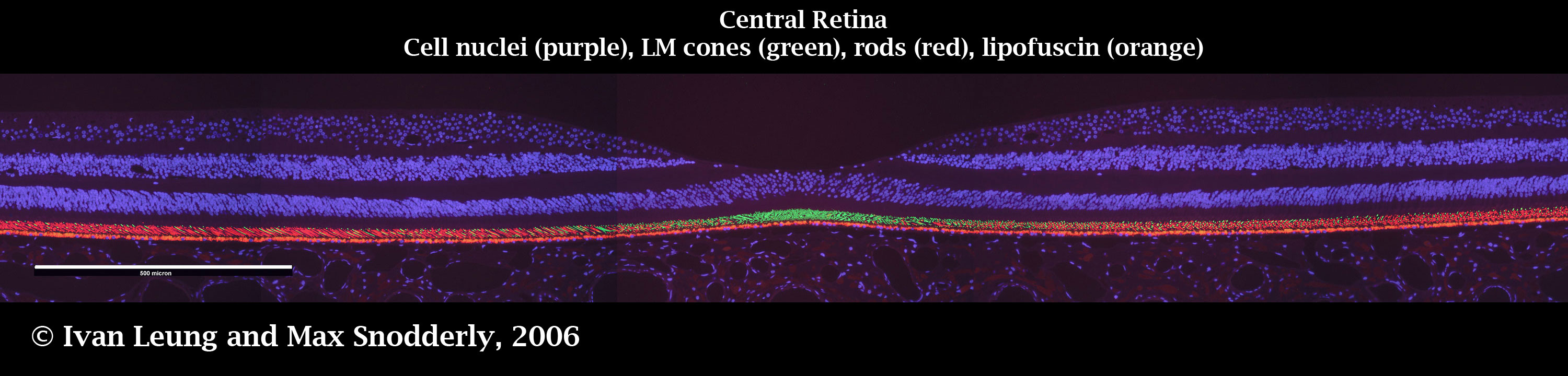
Fluorescence imaging to reveal four specific foveal features in one image through multiple exposures. Images of a two-micron-thick section along the vertical meridian were captured by epifluorescence microscopy at 400 x magnification. A composite of five microscope fields is shown here. Nuclei were stained with DAPI and visualized with a UV excitation filter set and appeared purple in color. Autofluorescence of the retinal pigment epithelium glows orange. Outer segments of the LM cones were labeled green with antibodies to LM cone opsin JH492 and images were captured with an FITC filter set. Outer segments of the rods were labeled red with an antibody to rhodopsin (Rho 4D2) and images were captured with a Texas Red filter set. Antibodies were generously supplied by Jeremy Nathans of Johns Hopkins University and R.S. Molday of the University of British Columbia. For further details see Leung, IY-F et al., Exp Eye Res. 2005; 81:513-529. Three separate images captured with UV, FITC and Texas Red filter sets were superimposed to visualize the features of the fovea and the distribution of LM cones and rods along the vertical meridian of the macula. This combined image illustrates vividly the small size of the area of high cone density. Note that the compression necessary to flatten the retina resulted in artifactual bending of the photoreceptors in the central fovea. Downloadable High Resolution Image
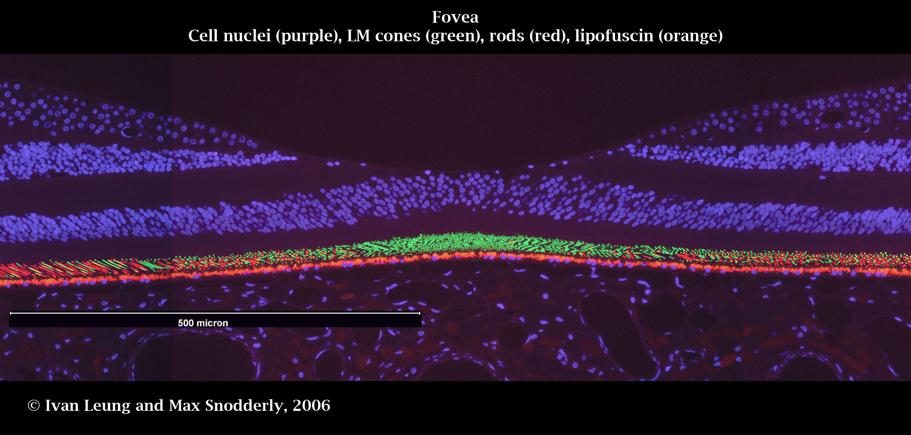
A composite of three microscope frames of the same retinal section provides more detail within the foveal depression. Downloadable High Resolution Image
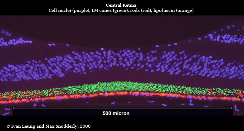
Devoting the whole image to one microscope frame of the central fovea provides the most detail we were able to attain. Downloadable High Resolution Image
Macular Pigment
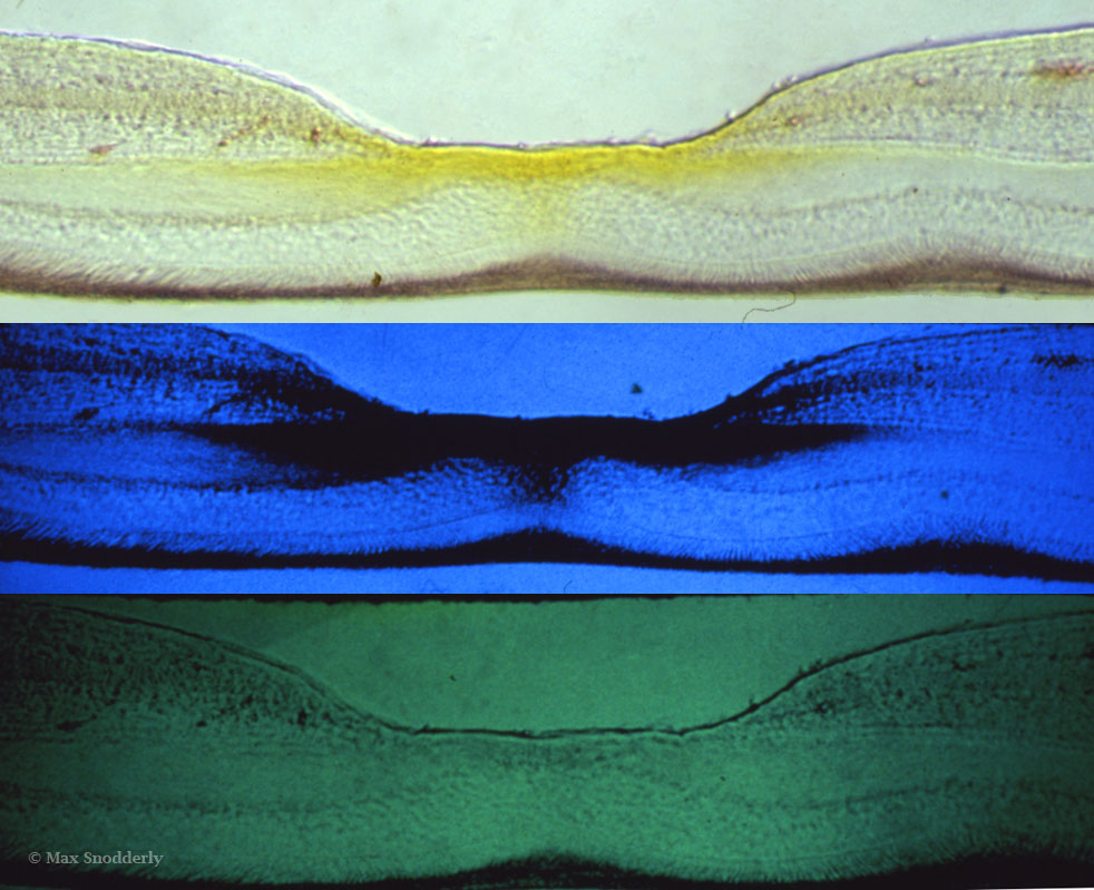
Frozen section through the fovea of a rhesus monkey viewed in white light, blue light, and green light. The yellow macular pigment appears dark in the middle panel because it absorbs blue light, but it is not visible in green light.
Downloadable High Resolution Image
Electrode Track Marking
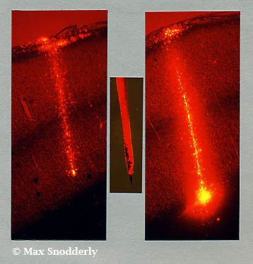
Fluorescence micrographs of marked electrode penetrations in primary visual cortex. The center panel shows the microelectrode coated with DiI except for the very tip. The penetration illustrated in the left panel reached layer 6, while the one in the right panel reached the white matter. From Snodderly and Gur (1995) J. Neurophysiol. 74: 2100-2125 (Cover Illustration).