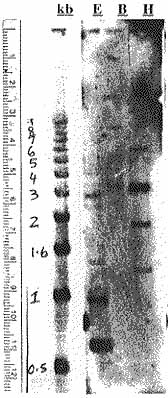
Figure 4. Southern hybridization blot with A. thaliana genomic DNA digested with EcoR1 (E), BamH1 (B), Hind III (H). Position and sizes ofmarkers present for comparison.

Figure 5. Northern hybridization blot with A. thaliana total RNA isolated from wounded seedlings, time 0 (t0), 1hr (t1), 5hrs (t5), 8hrs (t8), stems (S), floral tissue (F), leaves (L), and whole plant (W).