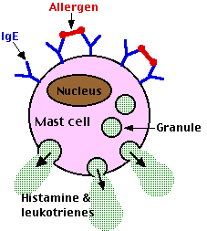
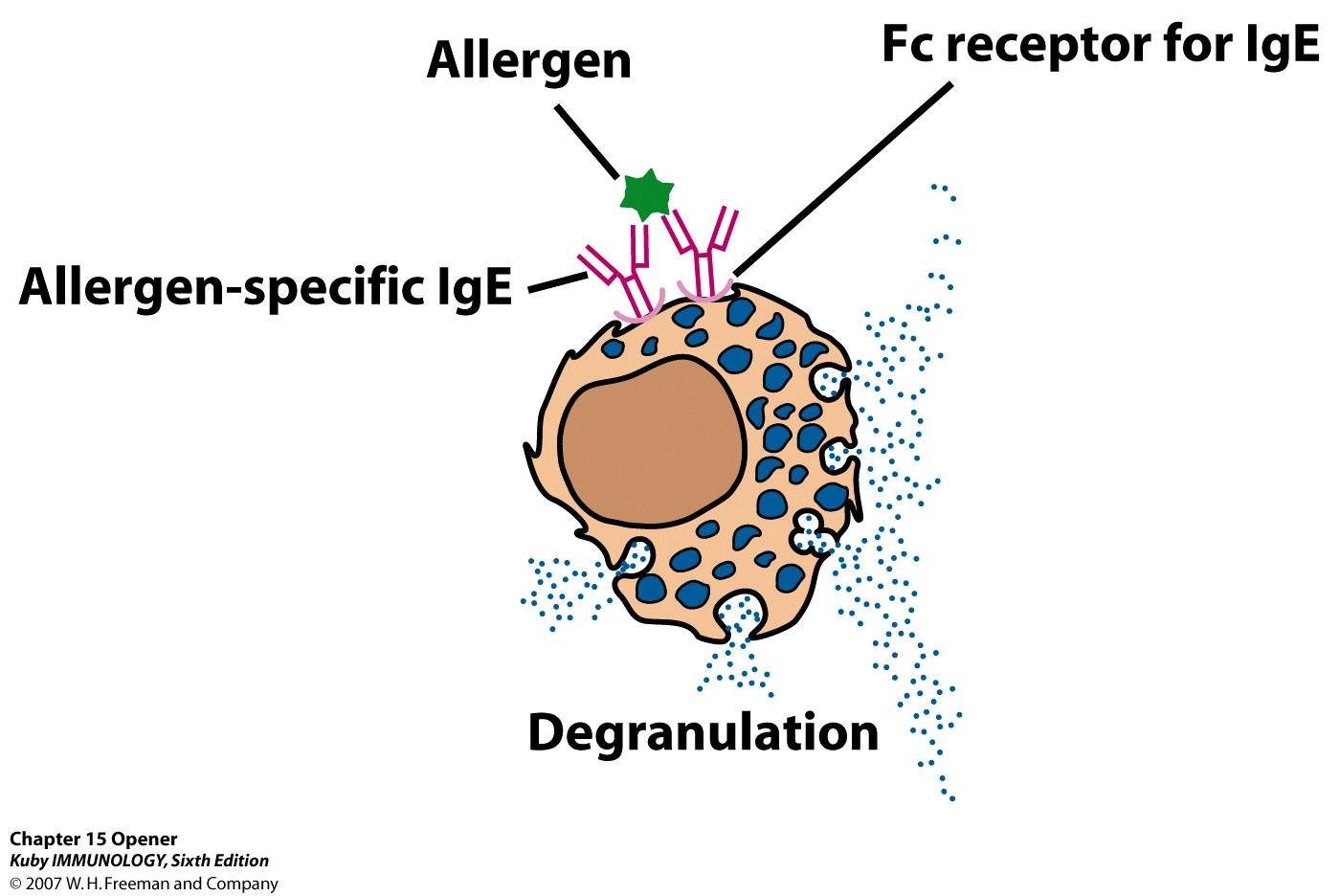
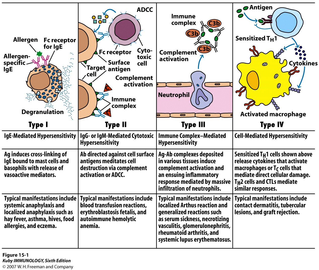
Hypersensitivity

There are four different types of hypersensitivities that result from different responses of the immune system:

Type I: Immediate hypersensitivity

- onset within minutes of antigen challenge

- examples are allergies to molds, insect bites

Type II: Cytotoxic hypersensitivity

- onset within minutes or a few hours of antigen challenge

- examples are adult hemolytic anemia and drug allergies

Type III: Immune complex-mediated hypersensitivity

- onset usually within 2-6 hours

- examples include serum sickness and systemic lupus

erythematosus

Type IV: Delayed Hypersensitivity

- inflammation by 2-6 hours; peaks by 24-48 hours

- examples include poison ivy and chronic asthma


There are 3 types of immediate hypersensitivities that depend on the interaction of antigens  with antibodies : Type I, Type II, and Type III.


Type I (IgE-mediated or anaphylactic-type) 

JAK/STAT pathway activation

These

Th2 Cells

Th2 cells are produced when  dendritic cells present antigen to the T cell's receptor for antigen (TCR) . The identity of the cytokine(s) is still uncertain (indicated by a ? in the figure).

The major lymphokines secreted by Th2 cells are


The Fc portion of IgE binds to the surface of mast cells and basophils

 When the allergen cross-links the Fab portions  of the mast cell-bound IgE, this triggers histamine  release by the mast cell, a process called degranulation, and the synthesis of other inflammatory mediators such as arachadonic acid, leukotrienes,  prostaglandins, and cytokines that contribute to inflammation (these act as chemoattractants). These agents cause the early phase of allergic reactions that appears within minutes after exposure to the antigen.

Late phase allergic reactions may begin several hours after exposure to antigen. It is thought that basophils play a major role here. Cell-bound IgE on the surface of basophils of sensitive individuals binds a substance called histamine releasing factor (possibly produced by macrophages and B-lymphocytes) causing further histamine release.

The inflammatory agents released or produced cause the following:

a. dilation  of blood vessels. This causes local redness (erythema) at the site of allergen delivery. If dilation is widespread, this can contribute to decreased vascular resistance, a drop in blood pressure, and shock .

b. increased capillary permeability. This causes swelling of local tissues (edema). If widespread, it can contribute to decreased blood volume and shock.

c. constriction of bronchial airways. This leads to wheezing and difficulty in breathing.

d. stimulation of mucous secretion. This leads to congestion of airways.

e. stimulation of nerve endings. This leads to itching and pain in the skin.

In a systemic anaphylaxis, the allergen is usually picked up by the blood and the reactions occur throughout the body. Examples include severe allergy to insect stings, drugs, and antisera. With a localized anaphylaxis, the allergen is usually found localized in the mucous membranes or the skin. Examples include allergy to hair, pollen, dust, dander, feathers, and food.

Type I hypersensitivity is treated symptomatically with such agents as:

a. epinephrine. Epinephrine relaxes smooth muscle, constricts blood vessels, and stimulates the heart. It is used for severe systemic reactions.

b. antihistamines . Antihistamines block the binding of histamine to histamine receptors on target cells.

c. Nasally administered steroids. Corticosteroids are potent antiinflammatory agents.

Severity may be reduced by desensitization shots (allergy shots). It is thought that when very dilute allergen is given by injection, it stimulates the production of IgG

IgG  then act as blocking antibodies to bind and neutralize much of the allergen in secretions before it can bind to the deeper cell-bound IgE on the mast cells in the connective tissue.

A new experimental approach to treating and preventing Type-I hypersensitivity involves giving the person with allergies injections of monoclonal antibodies  that have been made against the Fc  portion of human IgE.

This, in turn, blocks the attachment of the IgE to the Fc receptors on mast cells and basophils and the subsequent release of histamine by those cells upon exposure to allergen. In addition, the anti-IgE binds to IgE-producing B-lymphocytes causing apoptosis.

Q. how may mothers and children have the same allergic reactions?

IgG4 and IgE?


Type II (Antibody-dependent cytotoxicity) 

Mechanism: Either IgG or IgM is made against normal self antigens as a result of a failure in immune tolerance , or a foreign antigen resembling some molecule on the surface of host cells enters the body and IgG or IgM made against that antigen then cross reacts with the host cell surface. The binding of these antibodies to the surface of host cells then leads to:

a. opsonization  of the host cells whereby phagocytes stick to host cells by way of IgG,  and discharge their lysosomes  and ;

b. activation of the classical complement pathway causing MAC lysis  of the cells

c. ADCC  destruction of the host cells whereby NK cells  attach to the Fc portion  of the antibodies. The NK cell then release pore-forming proteins called perforins and proteolytic enzymes called granzymes. Granzymes pass through the pores and activate the enzymes that lead to apoptosis of the infected cell by means of destruction of its structural cytoskeleton proteins and by chromosomal degradation.


Type III (Immune complex-mediated) 

Mechanism: This is caused when soluble antigen-antibody (IgG or IgM) complexes, which are normally removed by macrophages in the spleen and liver, form in large amounts and overwhelm the body . These small complexes lodge in the capillaries, pass between the endothelial cells of blood vessels - especially those in the skin, joints, and kidneys - and become trapped on the surrounding basement membrane  beneath these cells . The antigen/antibody complexes then activate the classical complement pathway  . This may cause:

a. massive inflammation, due to complement protein C5a;

b. influx of neutrophils, due to complement protein C5a , resulting in neutrophils discharging their lysosomes and causing tissue destruction and furthes inflammation

c. MAC lysis  of surrounding tissue cells, due to the membrane attack complex, C5b6789n; and

d. aggregation of platelets, resulting in more inflammation and the formation of microthrombi that block capillaries.

This can lead to tissue death and hemorrhage.


Delayed Hypersensitivity (Type IV) 

Delayed hypersensitivity is cell-mediated rather than antibody-mediated.

Mechanism: Delayed hypersensitivity is the same mechanism as cell-mediated immunity. T8-lymphocytes  become sensitized to an antigen and differentiate into cytotoxic T-lymphocytes  while Th1 type T4-lymphocytes become sensitized to an antigen and produce cytokines . CTLs, cytokines, and/or macrophages then cause harm rather than benefit  .


Summary


ABO incompatibility

ABO incompatibility disease afflicts newborns whose mothers are blood type O, and who have a baby with type A, B, or AB.

Ordinarily, the antibodies against the foreign blood types A and B that circulate in mother's bloodstream remain there, because they are of a type that is too large to pass easily across the placenta into the fetal circulation. Some fetal red cells always leak into mother's circulation across the placental barrier (mother and fetal blood theoretically do not mix, but in actuality, they do to a small degree).

These fetal red cells stimulate the formation of a smaller type of anti-A or anti-B antibody which can pass into the baby's circulation and there cause the destruction of fetal red cells. The increased rate of destruction of red cells causes a subsequent increase in waste product production. This excess waste product, bilirubin, can overwhelm the normal waste elimination processes and lead to jaundice, the presence of excess bilirubin.