
![]() Histoplasmosis and Ajellomyces (Histoplasma)
capsulatum
Histoplasmosis and Ajellomyces (Histoplasma)
capsulatum
![]()
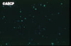
| Histoplasma capsulatum and Ajellomyces casulatus: in vitro | |||
 |
 |
 |
||||
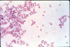
| Holoblastic macroconidia at 25°C | Hypha and tuberculate macroconidia | Yeast cells at 37°C | ||||
 |
||||||
|
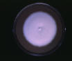
Typical older white mold colony at 25°C
Older = after many subcultures |
||||||
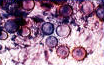
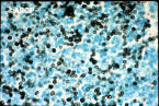
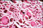
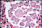
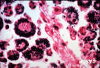
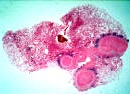
| H. capsulatum: In tissue | |||
 |
 |
 |
||||
| In liver | ||||||
 |
 |
 |
||||
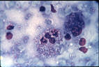
| mΦ with many yeast cells from acute pulmonary case | ||||||
 |
||||||
| H + E; yeast cells in phagocyte yeast cell shrunk from cell wall looks like a capsule | ||||||
| Histoplasmosis: | Residual lesions | ||
 |
 |
 |
||||
| Residual calcified lesions in lung | Lesions (calcified) in spleen from previous acute histoplasmosis (0.2-0.4 cm in diameter) | |||||
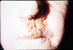
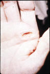
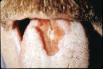
| Cutaneous lesions: | |||
 |
 |
 |
||||
| From dissemination | From dissemination | From dissemination | ||||
 |
||||||
| Primary lesion from lab accident | ||||||
|
|
|||||