
![]() Dermatophyte and
Dermatophytosis
Dermatophyte and
Dermatophytosis
![]()
Tinea barbae / Tinea capitus: Prepubertal / Tinea capitus: Black dot /
Tinea favosa: Favus / Arthrodermas gypseum & Microsporum gypseum /
Tinea corporis / Tinea cruris / Tinea imbricata / Tinea manuum / Tinea pedis /
 |
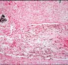
||
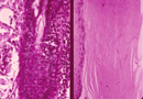
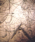
| (right) Dermatophyte infection. The hyphal strands penetrate through the stratum corneum but do not invade the living cells of the epidermis. (left) Mycelium growing down a hair shaft to the bulb (Periodic acid--Schiff stain X400) |
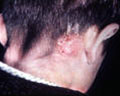
| Tinea barbae: Ringworm of facial hair | |||
 |
 |
 |
||||
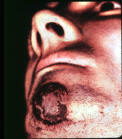
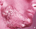
| Topical, mild form | Pustular & abscessed form | Deep pustule folliailitus by T. verrucosum |
|
|
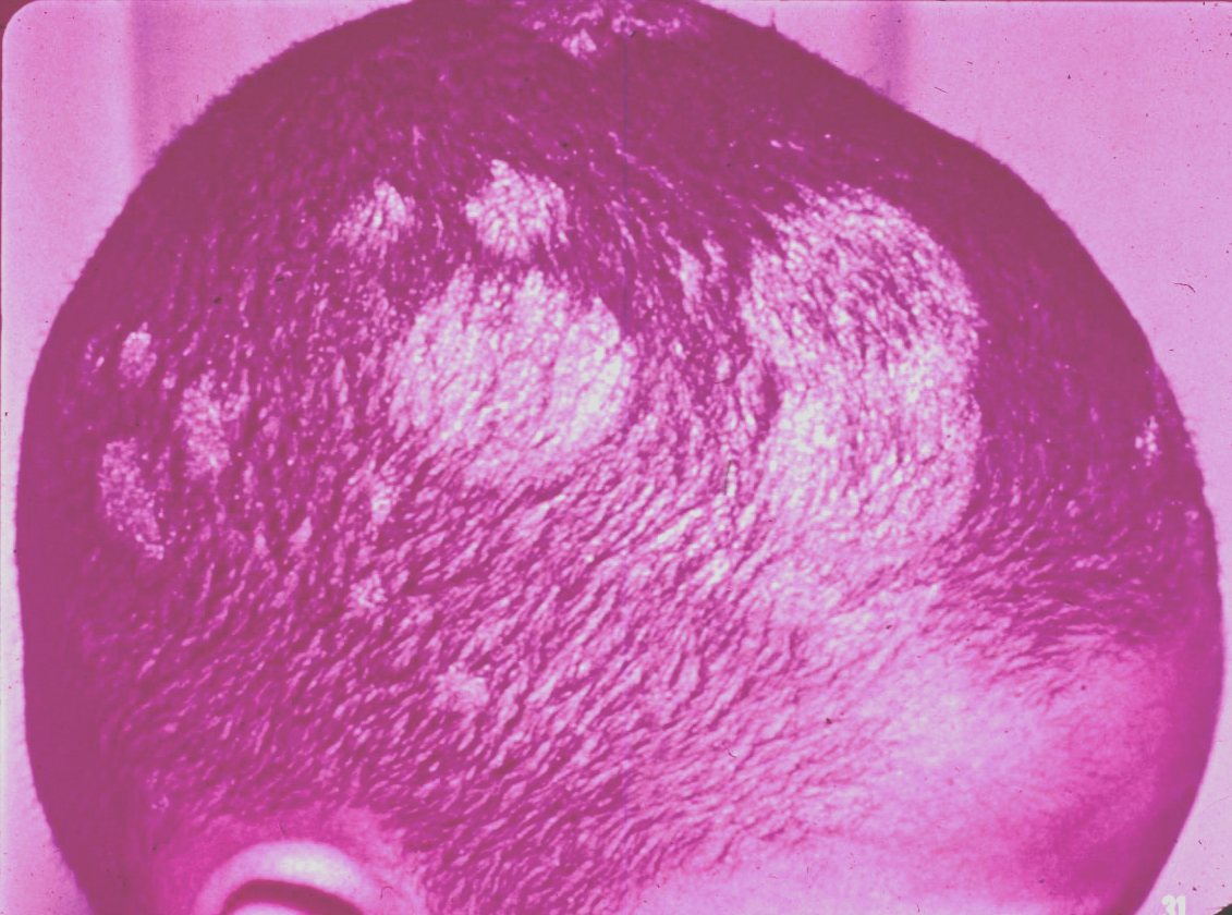
| Tinea capitus: Prepubertal, gray patch, classical scalp ringworm, and epidemic ringworms. | |||
 |
 |
 |
||||
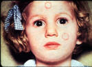
| Caused by M. audouinii | Cuased by M. audouinii | Caused by unidentified Microsporum (Probably M. gypsium or M. canis because pustular and suppurative) | ||||
 |
 |
 |
||||
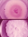
| Caused by M. canis | M. canis on SABS (reverse and top), agent of gray patch | M. canis macroconidia, agent of gray patch |
 |
 |
|||||
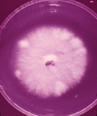
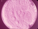
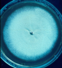
| M. audouinii on SDA, colony with pleomerphic sectors | M. gypseum, agent of gray patch | |||||
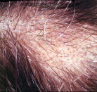
| Tinea capitus: Black dot ringworm | |||
 |
 |
 |
||||
| By T. violaceum | T. violaceum | T. tonsurans | ||||
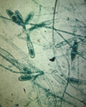
 |
 |
|||||
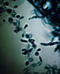
| T. tonsurans (micro conidia in vitro) | T. tonsurans (micro and macro conidia) |
|
|
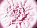
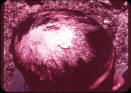
| Tinea Favosa: Favus, a special kind of tinea capitus | |||
 |
 |
 |
||||
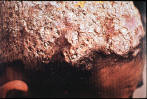
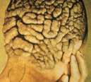
| Seborrheic stage showing matted hair (infected hair is grey) | Advanced disease with alopecia and scarring | Massive crust of scutula, mycilium and scalp epithelium | ||||
 |
 |
 |
||||
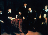
| T. schoenleinii | Ferdinand Bol's painting, "Four Governors of the Amsterdam Leper Asylum" | Trichophyton schoenleinii, cup-shaped scutula |
|
|
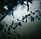
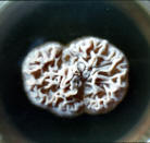
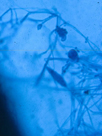
| Arthrodermas gypseum: Telomorph | ||
| Microsporum gypseum: Anamorph |
 |
 |
 |
||||
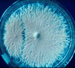
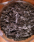
| On PDA (anamorph) | Mated strains in hair-soil culture | Gymnothecia on hair | ||||
 |
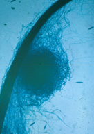 |
 |
||||
| Antheridia, ascogonia, and conidium | Gymnothecia on hair | Asci and ascospores (Asci prototunicate) | ||||
|
|
|
 |
||||
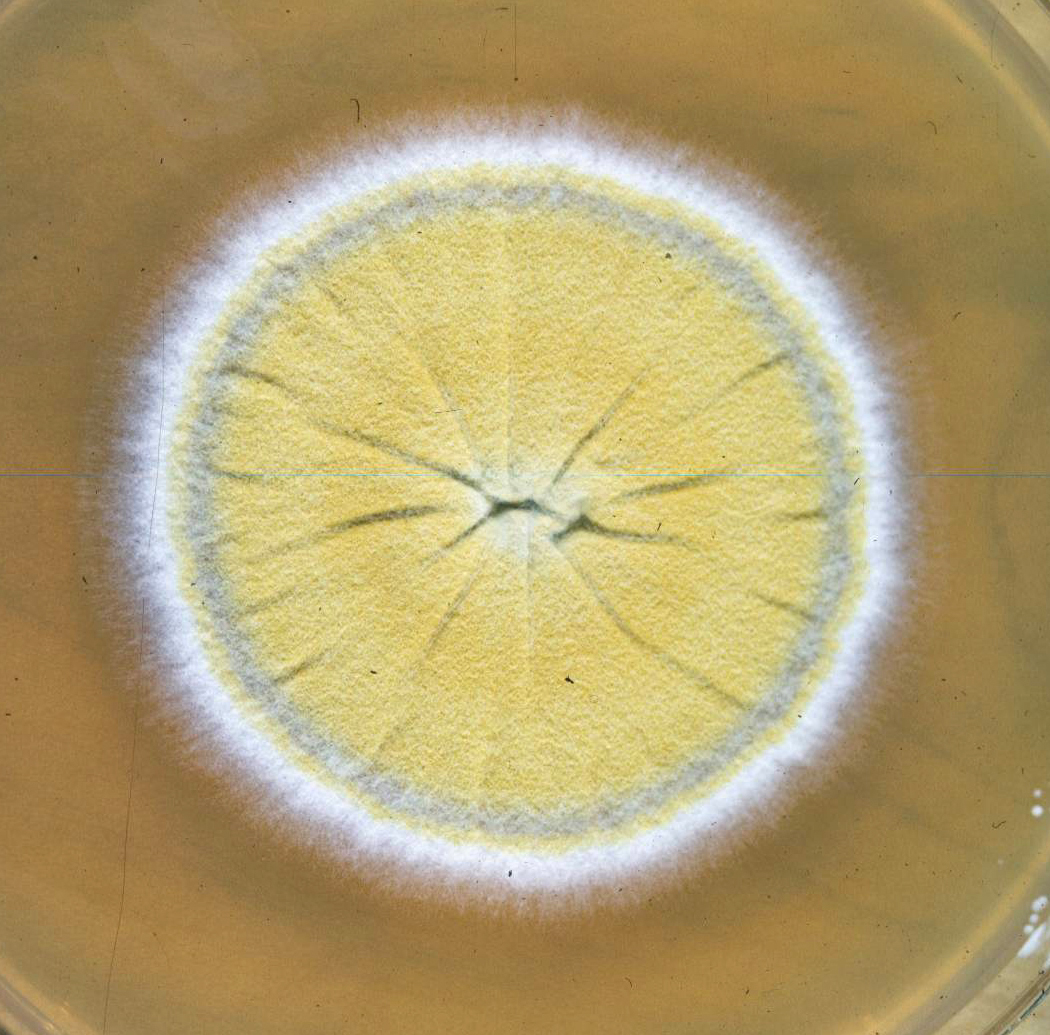
| Microsporum canis (reverse and top) | M. canis holothallic conidia | Epidermophyton floccosum on PDA | ||||
 |
 |
 |
||||
| E. floccosum holothallic conidia | Epidermophyton floccosum on PDA | T. tonsurans holoarthric conidia |
|
|
| Tinea corporis: | |||
 |
 |
|||
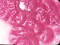
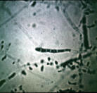
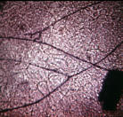
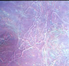
| Infected skin scales & Parkers ink (bright field) | Treated with KOH (Phase-contrast) | |||
 |
 |
|||
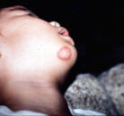
| Lesion on chin (non-vesicular form) | Lesions on forhead & cheek (Non-vesicular form) |
|
|
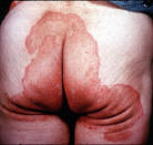
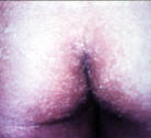
| Tinea cruris: Ringworm of groin and periannal regions | |||
 |
 |
|||
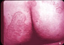
| Annular type of gluteal areas | Annular type on lower trunk | |||
 |
 |
|||
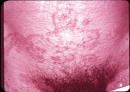
| Periannal type | Periannal type |
|
|
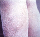
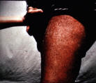
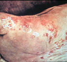
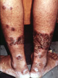
| Tinea imbricata: Beauty ringworm | |||
 |
 |
|||
| Concentric rings over chest (Free edges of scales face inward) | Chronic scaling infection on legs of female |
 |
 |
|||
| Severe remission case on legs of male | T. concentricium |
|
|
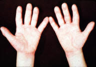
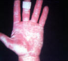
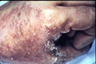
| Tinea manuum: | |||
 |
 |
 |
||||
| By T. rubrum | By T. rubrum | By T. rubrum |
|
|
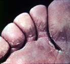
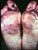
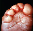
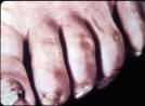
| Tinea pedis: Athlete's foot | |||
 |
 |
 |
||||
| Interdigital | Hyperkeratotic & erythrodermic | Mild children's form | ||||
 |
 |
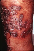 |
||||
| Vesicular form | Deep hyperkeratotic form involving feet, toenails and legs | Caused by T. mentagrophytes | ||||
 |
|
|
||||
| Dermatophytic pseudo-mycetoma caused by M. canis |
|
|
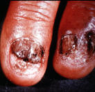
| Tinea unguium: Dermatophytic onychomycosis | |||
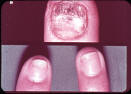 |
 |
|||
| (top) Advanced disease; (bottom) Infection at distal edges of nail plate | Groved hyperkerototic nails | |||
 |
 |
|||
| Toenail infections by T. rubrum | T. rubrum |
|
|
|||||