 Chapter
9. Secretory
cells and tissues.
Chapter
9. Secretory
cells and tissues.
The figures presented here were selected to illustrate aspects of secretory structures in plants, and the legends were written to complement the more complete discussion of secretion presented in Chapter 9 (pages 141 to 166) in the textbook Plant Anatomy by J. D. Mauseth, published by Cummings & Hathaway. That chapter has several diagrams that may be especially helpful in understanding some of the micrographs presented here. This web site will present only the types of secretory structures you are likely to encounter in a teaching laboratory of Plant Anatomy, so electron micrographs are not presented; the textbook does provide those.
Click here for a set of links to the micrographs of this chapter.
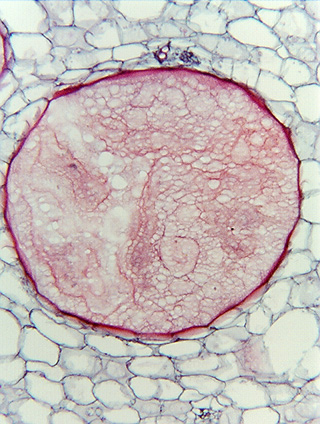
Mucilage secreting cell of the cactus Acanthocereus columbianus.