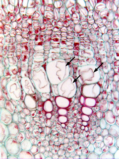
 Fig.
7.5-4a.
Transverse section of developing vessels in roots of oak (Quercus). This
bundle contains many mature vessel elements, readily identifiable by their large
diameter and red-stained, lignified secondary walls. But the cells marked with
arrows have the proper size to be vessel elements but they do not have secondary
walls. Also, there is a bit of plasmolyzed protoplasm in them. These
are vessel elements that are still differentiating – they appear to
have finished their enlargement and had not even begun to deposit the S1
layer of the secondary wall. It is not too common to see vessel elements in this
immature stage of development, they seem to pass through this stage quickly.
Fig.
7.5-4a.
Transverse section of developing vessels in roots of oak (Quercus). This
bundle contains many mature vessel elements, readily identifiable by their large
diameter and red-stained, lignified secondary walls. But the cells marked with
arrows have the proper size to be vessel elements but they do not have secondary
walls. Also, there is a bit of plasmolyzed protoplasm in them. These
are vessel elements that are still differentiating – they appear to
have finished their enlargement and had not even begun to deposit the S1
layer of the secondary wall. It is not too common to see vessel elements in this
immature stage of development, they seem to pass through this stage quickly.