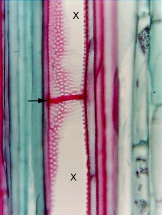
 Fig.
7.3-6c. Longitudinal section of vascular bundle of
corn. The microtome knife in this section cut away all the front wall of the
vessel and just grazed the back wall, cutting away much of it from the lower
vessel element (at X) and about half of it from the upper element (X). All the
back wall is present where the two vessel elements meet at the perforation, and
the perforation plate rim is visible as a dark red band at the arrow. Note there
is a fine black line that runs horizontally through the rim, indicating that
this is really two structures, the perforation plate of the upper cell glued to
that of the lower cell by the middle lamella. Where the back wall is present, we
can see that it has rather irregular pitting, some of the pits being circular,
others more elongate, almost scalariform. Also, the pitting occurs even very
close to the perforation plate itself.
Fig.
7.3-6c. Longitudinal section of vascular bundle of
corn. The microtome knife in this section cut away all the front wall of the
vessel and just grazed the back wall, cutting away much of it from the lower
vessel element (at X) and about half of it from the upper element (X). All the
back wall is present where the two vessel elements meet at the perforation, and
the perforation plate rim is visible as a dark red band at the arrow. Note there
is a fine black line that runs horizontally through the rim, indicating that
this is really two structures, the perforation plate of the upper cell glued to
that of the lower cell by the middle lamella. Where the back wall is present, we
can see that it has rather irregular pitting, some of the pits being circular,
others more elongate, almost scalariform. Also, the pitting occurs even very
close to the perforation plate itself.