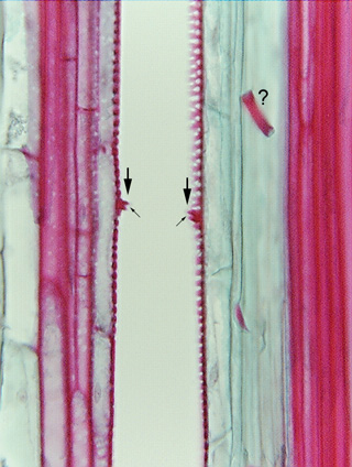
 Fig.
7.3-6a. Longitudinal section of vascular bundle in
corn. (Zea mays). The microtome knife passed down the back of this
vessel, removing the back wall, then on the next cut, removed the front wall.
All we can see are the two side walls. The arrows indicate the rims
of a perforation plate, and the open area between the two arrow tips
is the perforation itself. Remember, there are actually two perforation plates
here, one for the lower cell, one for the upper one; if your computer screen is
good, you may be able to see a slight indentation (small arrows) that shows it
is a double structure. In some species, the rims are so narrow they are almost
impossible to see; in other species they would project into the vessel lumen a
little farther than this.
What is the red band in the upper right corner?