 Fig.
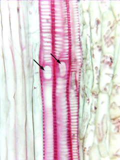
7.3-5a. Longitudinal section of vascular bundles
in spurge (Euphorbia). There are three vessels oriented vertically in the
center of this micrograph, and two
have simple, round perforations facing us (marked by arrows). All
three vessels have pits that are predominantly scalariform, but notice that
there is a bit of pit-free wall immediately surrounding each perforation: that
pit-free area is the perforation plate rim. Take another look at the two
perforations and keep in mind that is where two separate vessel elements contact
each other. Although each perforation appears to just be a hole in a secondary
wall, it actually consists of two perforations that are so perfectly aligned we
cannot see there are two. Rarely, there will be slight misalignment and the dual
nature will be visible, but usually the high magnification and resolution of
scanning electron microscopy is needed to see that.
Fig.
7.3-5a. Longitudinal section of vascular bundles
in spurge (Euphorbia). There are three vessels oriented vertically in the
center of this micrograph, and two
have simple, round perforations facing us (marked by arrows). All
three vessels have pits that are predominantly scalariform, but notice that
there is a bit of pit-free wall immediately surrounding each perforation: that
pit-free area is the perforation plate rim. Take another look at the two
perforations and keep in mind that is where two separate vessel elements contact
each other. Although each perforation appears to just be a hole in a secondary
wall, it actually consists of two perforations that are so perfectly aligned we
cannot see there are two. Rarely, there will be slight misalignment and the dual
nature will be visible, but usually the high magnification and resolution of
scanning electron microscopy is needed to see that.