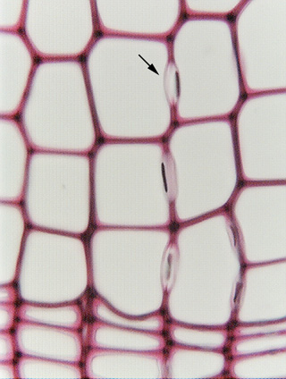
 Fig.
7.2-11. Transverse section of pine wood. This high
magnification view of pine tracheids shows several circular
bordered pits cut in transverse section (they are shown in face view
in Fig. 7.2-6). The pale pink region that arcs into the tracheid lumen is the
pit border (compare this view with the diagram Fig 7.10 and the micrograph 7.12
on pages 116 and 117 in Plant Anatomy [Mauseth]). The dark, almost black
bar is the torus; in all these pits, the torus is pressed against one side of
the pit. Having the torus to one side like this indicates that the wood had
suffered from water stress and the water columns had cavitated (broken); this
sample was probably taken from heartwood, or maybe even just from a piece of dry
lumber. When the water rushed from the cavitating cell to the still functional
one, the rapid flow pushed the torus against the inner aperture where it became
stuck. The displacement of the torus seals the pit and prevents air from passing
into the water-filled tracheid, thus preventing the embolism from spreading.
Fig.
7.2-11. Transverse section of pine wood. This high
magnification view of pine tracheids shows several circular
bordered pits cut in transverse section (they are shown in face view
in Fig. 7.2-6). The pale pink region that arcs into the tracheid lumen is the
pit border (compare this view with the diagram Fig 7.10 and the micrograph 7.12
on pages 116 and 117 in Plant Anatomy [Mauseth]). The dark, almost black
bar is the torus; in all these pits, the torus is pressed against one side of
the pit. Having the torus to one side like this indicates that the wood had
suffered from water stress and the water columns had cavitated (broken); this
sample was probably taken from heartwood, or maybe even just from a piece of dry
lumber. When the water rushed from the cavitating cell to the still functional
one, the rapid flow pushed the torus against the inner aperture where it became
stuck. The displacement of the torus seals the pit and prevents air from passing
into the water-filled tracheid, thus preventing the embolism from spreading.
In most Plant Anatomy Laboratories, you will see wood like this. Occasionally, the pine wood sample will have been taken from still-functional sapwood, and care will have been take to let the wood dry slowly, without abrupt cavitation. In such samples, you may see the torus suspended between the two halves of the pit-pair, rather than stuck to one side.