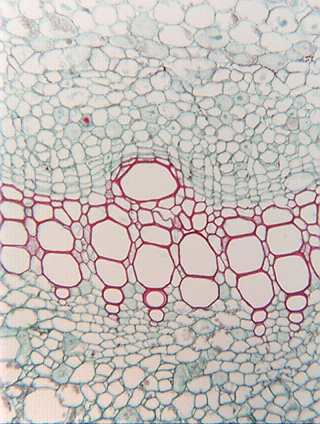
 Fig.
7.1-1. Transverse section of sweet potato
stem (Ipomoea batatas). This micrograph is intended as an
introduction to xylem. In almost all histological slides, xylem will
be stained red because of its thick, lignified walls. Many or most xylem cells
will be much larger than nearby cells. These are vessel elements in transverse
section; because they are mature, they have no protoplasts. They may have been
filled with water being conducted when this sampled was dissected from the
plant, but the water either drained out during dissection or it was removed by
dehydration. You will never see slides of xylem with their water actually in
them unless the material was frozen before dissection and then kept frozen until
you examine it – something which is extremely difficult to do.
Fig.
7.1-1. Transverse section of sweet potato
stem (Ipomoea batatas). This micrograph is intended as an
introduction to xylem. In almost all histological slides, xylem will
be stained red because of its thick, lignified walls. Many or most xylem cells
will be much larger than nearby cells. These are vessel elements in transverse
section; because they are mature, they have no protoplasts. They may have been
filled with water being conducted when this sampled was dissected from the
plant, but the water either drained out during dissection or it was removed by
dehydration. You will never see slides of xylem with their water actually in
them unless the material was frozen before dissection and then kept frozen until
you examine it – something which is extremely difficult to do.