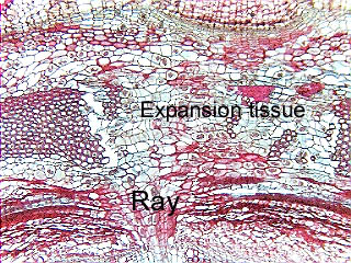
 Fig.
16.3-2a and b. Transverse section of Dutchman’s pipe (also called
birthwort; Aristolochia). This is a young stem with only a small amount
of secondary phloem (barely visible in the low magnification view), but you can
see that the cortex has thick bands of fibers (arrows). The formation of
secondary xylem and phloem has pushed the cortex and its fibers outward, and
rather than rupturing, dilatation tissue has formed.
There were parenchyma cells among the fibers, and they have divided and enlarged
in the region marked by the double-headed arrow (shown in the high magnification
view). Because this is dilatation by isolated parenchyma cells in the cortex,
it is expansion tissue. The ray itself is also dilatating with
proliferative tissue.
Fig.
16.3-2a and b. Transverse section of Dutchman’s pipe (also called
birthwort; Aristolochia). This is a young stem with only a small amount
of secondary phloem (barely visible in the low magnification view), but you can
see that the cortex has thick bands of fibers (arrows). The formation of
secondary xylem and phloem has pushed the cortex and its fibers outward, and
rather than rupturing, dilatation tissue has formed.
There were parenchyma cells among the fibers, and they have divided and enlarged
in the region marked by the double-headed arrow (shown in the high magnification
view). Because this is dilatation by isolated parenchyma cells in the cortex,
it is expansion tissue. The ray itself is also dilatating with
proliferative tissue.
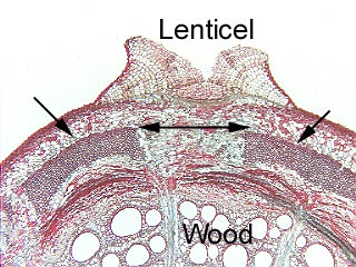
Notice that
the alignment of the ray, the expansion tissue and the lenticel: the
lenticel permits oxygen to diffuse into the stem, and the expansion tissue has
created an aerenchymatous path through the fiber band (an intact fiber band
would hinder oxygen diffusion). The dilatation of the ray will also create more
intercellular spaces that will aid oxygen movement.