 Fig.
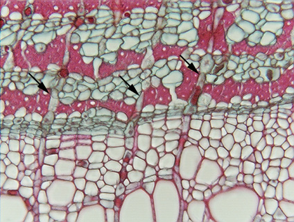
16.2-1a and b. Transverse section of linden (Tilia). The low
power micrograph gives a general orientation, with secondary xylem at the
bottom, phloem at the top, and the vascular cambium running horizontally across
the center (indicated by arrows). This
secondary phloem is more complex than that of pine. The bands that
are stained dark red consist of secondary phloem fibers, seen more clearly in
the high power view. Alternating with the bands of fibers are bands consisting
predominantly of sieve tube members and companion cells. It is tempting to
assume that these phloem bands correspond to the annual rings in xylem, but
typically they are not annual: several sets can be produced per year.
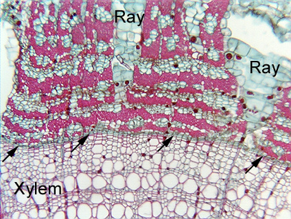
Fig.
16.2-1a and b. Transverse section of linden (Tilia). The low
power micrograph gives a general orientation, with secondary xylem at the
bottom, phloem at the top, and the vascular cambium running horizontally across
the center (indicated by arrows). This
secondary phloem is more complex than that of pine. The bands that
are stained dark red consist of secondary phloem fibers, seen more clearly in
the high power view. Alternating with the bands of fibers are bands consisting
predominantly of sieve tube members and companion cells. It is tempting to
assume that these phloem bands correspond to the annual rings in xylem, but
typically they are not annual: several sets can be produced per year.
Two
rays are visible in the phloem, and are a bit unusual in being
narrower near the cambium (where they are young) and wider away from the cambium
(where they are older; remember that phloem is pushed outward by formation of
new phloem interior to pre-existing phloem). The width of the rays results
because ray cells themselves divide (a process called dilatation and illustrated
in micrographs below).