|
[Home]
[Up]
| |
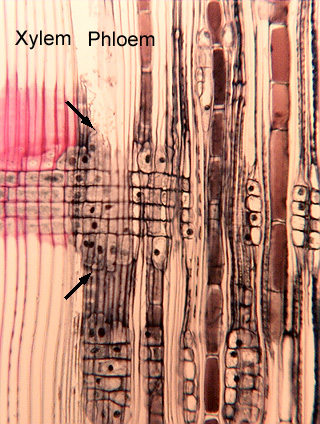 Fig.
16.1-4.
Radial section of secondary phloem of pine. This radial section shows xylem on
the left and phloem on the right; between the two labels is the vascular
cambium. Examine
the ray between the two arrows. In the vascular cambium at this point
is a set of ray initials that has produced the xylem ray (with ray tracheids
visible along the top) as well as the phloem ray (in the phloem, the uppermost
cells differentiate as tall parenchyma cells, not as ray tracheids). This
section shows only a little of the phloem ray; why? Notice the stacks
of parenchyma cells on the right side of the micrograph, aligned with the ray
– those are part of the same ray. As the sieve cells collapse, the ray is
thrown into undulations, so a straight cut with the microtome knife will not be
able to follow the undulating ray. The section ends up having just short
segments of the ray. Fig.
16.1-4.
Radial section of secondary phloem of pine. This radial section shows xylem on
the left and phloem on the right; between the two labels is the vascular
cambium. Examine
the ray between the two arrows. In the vascular cambium at this point
is a set of ray initials that has produced the xylem ray (with ray tracheids
visible along the top) as well as the phloem ray (in the phloem, the uppermost
cells differentiate as tall parenchyma cells, not as ray tracheids). This
section shows only a little of the phloem ray; why? Notice the stacks
of parenchyma cells on the right side of the micrograph, aligned with the ray
– those are part of the same ray. As the sieve cells collapse, the ray is
thrown into undulations, so a straight cut with the microtome knife will not be
able to follow the undulating ray. The section ends up having just short
segments of the ray.
|