 Fig.
15.2-5a and b. Radial section of white pine wood. The lowest row of
ray cells consists of ray
tracheids. These can be difficult to pick out, and when examining
them with your microscope, focus up and down frequently at high power to pick
out details. Look for the circular bordered pits on the end walls (indicated by
arrows here) as well as small circular bordered pits on the radial walls (the
small white areas that are out of focus here).
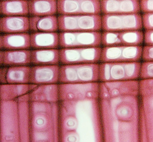
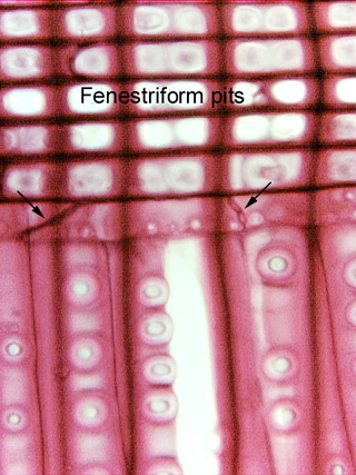
Fig.
15.2-5a and b. Radial section of white pine wood. The lowest row of
ray cells consists of ray
tracheids. These can be difficult to pick out, and when examining
them with your microscope, focus up and down frequently at high power to pick
out details. Look for the circular bordered pits on the end walls (indicated by
arrows here) as well as small circular bordered pits on the radial walls (the
small white areas that are out of focus here).
Above the row of ray tracheids are
rows of ray parenchyma cells with large fenestriform
(window-like) pits. The fenestriform pits are a type of cross-field
pitting: pitting that interconnects ray cells with axial cells. The
lower micrograph was refocused to bring the fenestriform pits into sharper view.
You can see one or two fenestriform pits in each cell, the pits occupying almost
the entire wall.