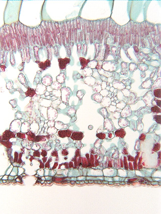
 Fig.
10.6-2. Fig leaf. This is a magnification of the
lower portion of a fig leaf; the lower
epidermis appears to be uniseriate, but immediately interior to it
are two layers of large, empty-looking cells. Without being able to examine
young leaves in an early stage of development, we cannot be certain.
Fig.
10.6-2. Fig leaf. This is a magnification of the
lower portion of a fig leaf; the lower
epidermis appears to be uniseriate, but immediately interior to it
are two layers of large, empty-looking cells. Without being able to examine
young leaves in an early stage of development, we cannot be certain.
A stoma is present.