 Fig.
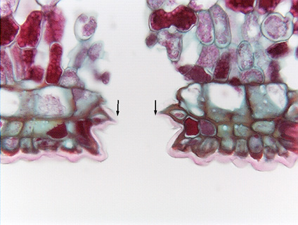
10.3-8. Magnification of fig leaf stoma. This micrograph shows you a common artifact: the stoma appears to have a very wide
stomatal pore. In fact, this pore is too wide for the guard cells to be able to
close it, no matter how much they shrink. But this
view reflects an error rather than the reality of what the stoma was like on the
plant: between the time the section was cut and when it was glued
onto the microscope slide, it expanded, and the guard cell on one side of the
pore was pulled far away from the guard cell on the other side. This width is so
extraordinary that it arouses our suspicion, but if it had pulled only slightly
apart, we might not have suspected an artifact. Even in sections that appear to
be perfect, it would be risky to try to measure the width of the pore – we
could never be certain we were not measuring artifacts.
Fig.
10.3-8. Magnification of fig leaf stoma. This micrograph shows you a common artifact: the stoma appears to have a very wide
stomatal pore. In fact, this pore is too wide for the guard cells to be able to
close it, no matter how much they shrink. But this
view reflects an error rather than the reality of what the stoma was like on the
plant: between the time the section was cut and when it was glued
onto the microscope slide, it expanded, and the guard cell on one side of the
pore was pulled far away from the guard cell on the other side. This width is so
extraordinary that it arouses our suspicion, but if it had pulled only slightly
apart, we might not have suspected an artifact. Even in sections that appear to
be perfect, it would be risky to try to measure the width of the pore – we
could never be certain we were not measuring artifacts.