 Fig.
10.3-3.
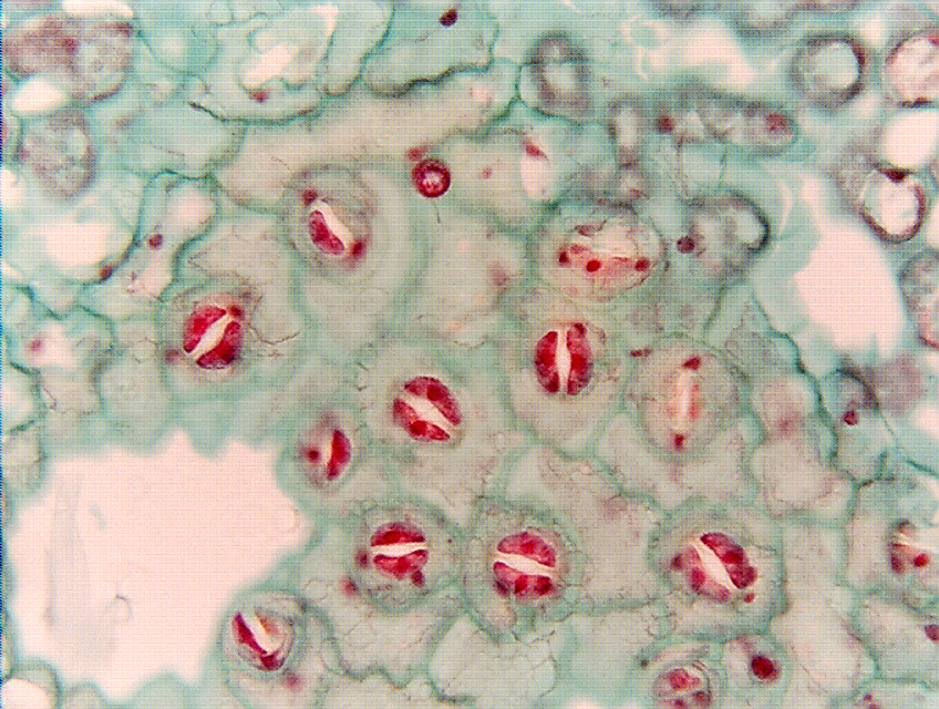
Paradermal section of leaf of ground ivy (Glechoma; in the mint family,
not a real ivy). A
paradermal section is one in which the microtome knife cuts parallel
to the surface of an organ, parallel to the surface of the leaf in this case. If
the leaf were perfectly flat, the knife might cut a uniform section from one end
to the other of the leaf, and extending uniformly to both sides. BUT,
leaves tend to be curved or undulate, so the knife cuts into part of the leaf
and misses other parts on the first pass. In this section, the knife has just
cut into the epidermis in the center of the micrograph (where all the stomata
are visible), and missed it at the two white spots (the leaf was curved away
from the plane of sectioning there). Notice that the guard cells are very
distinct but the surrounding ordinary epidermis cells are not: the guard cells
must be a bit higher or lower than the ordinary epidermis cells. The few cells
with dots are mesophyll (the dots are chloroplasts): those parts of the leaf are
curved up toward the plane of sectioning and their epidermis is in the section
that was cut before this one.
Fig.
10.3-3.
Paradermal section of leaf of ground ivy (Glechoma; in the mint family,
not a real ivy). A
paradermal section is one in which the microtome knife cuts parallel
to the surface of an organ, parallel to the surface of the leaf in this case. If
the leaf were perfectly flat, the knife might cut a uniform section from one end
to the other of the leaf, and extending uniformly to both sides. BUT,
leaves tend to be curved or undulate, so the knife cuts into part of the leaf
and misses other parts on the first pass. In this section, the knife has just
cut into the epidermis in the center of the micrograph (where all the stomata
are visible), and missed it at the two white spots (the leaf was curved away
from the plane of sectioning there). Notice that the guard cells are very
distinct but the surrounding ordinary epidermis cells are not: the guard cells
must be a bit higher or lower than the ordinary epidermis cells. The few cells
with dots are mesophyll (the dots are chloroplasts): those parts of the leaf are
curved up toward the plane of sectioning and their epidermis is in the section
that was cut before this one.