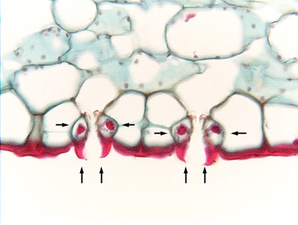
 Fig.
10.3-18. Transverse section of leaf of kafir-lily (Clivia, an
ornamental monocot). Examine the guard cells (horizontal arrows) and their outer
ledges (vertical arrows): the cuticle on the guard cells differs from
that over all the rest of the leaf, having accumulated to a tremendous
thickness. When guard cells lose turgor and shrink, they move toward each other,
closing the stomatal pore as the facing walls of the guard cells push against
each other. But in species that have ledges like this, the
ledges are the first region to make contact and seal the pore, then
the facing walls push against each other: there are actually two seals that keep
water from squeezing out through a closed stoma. Some species even have inner
ledges also, providing a third seal.
Fig.
10.3-18. Transverse section of leaf of kafir-lily (Clivia, an
ornamental monocot). Examine the guard cells (horizontal arrows) and their outer
ledges (vertical arrows): the cuticle on the guard cells differs from
that over all the rest of the leaf, having accumulated to a tremendous
thickness. When guard cells lose turgor and shrink, they move toward each other,
closing the stomatal pore as the facing walls of the guard cells push against
each other. But in species that have ledges like this, the
ledges are the first region to make contact and seal the pore, then
the facing walls push against each other: there are actually two seals that keep
water from squeezing out through a closed stoma. Some species even have inner
ledges also, providing a third seal.
Even when the stomatal pore is open, the ledges help retain water by acting somewhat like the grooves of yucca leaves or the overarching subsidiary cells of Hakea: because the ledges are so tall, they create a miniature tube with the stomatal pore at one end.
The guard cells are much smaller -- both narrower and thinner -- than the surrounding epidermis cells (from this view, we cannot tell if they are ordinary epidermis cells or subsidiary cells). (Dark red bodies in the guard cells are nuclei; the dots in the mesophyll cells are chloroplasts.)