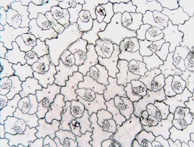
Fig. 10.3-1. Epidermal peel of sedum (Sedum). These preparations were made by grasping the epidermis with fine forceps, then slowly peeling the epidermis away from the leaf. In the low magnification, notice the sinuous, wavy walls of the large cells: that is a sure sign that this is from a dicot. All those cells with sinuous walls are ordinary epidermal cells. The small clusters of cells are stomatal complexes. The low magnification view shows that the stomatal complexes are located close together in this species, with only one or two ordinary epidermal cells between each complex.
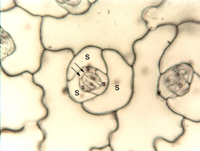
The high magnification view shows a stomatal complex. Even at this high magnification, the two guard cells are small (arrows), and the stomatal pore is closed and not visible. In addition, the stomatal complexes contain subsidiary cells (marked by S), which can be identified by their non-sinuous walls and crescent shape.