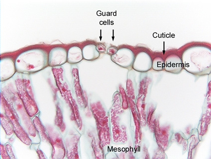
 Fig.
10.2-2. Transverse section of leaf of Peucephyllum (no common
name). Most epidermis cells visible here are ordinary
epidermis cells, but there are also two guard cells and the stomatal pore
located between them. Note that the stomatal pore is the only intercellular
space in the epidermis. The outer wall of each epidermis cell bulges outward due
to turgor pressure, and the cuticle follows the contours of the cells, being
curved itself. This cuticle is unusually thick.
Fig.
10.2-2. Transverse section of leaf of Peucephyllum (no common
name). Most epidermis cells visible here are ordinary
epidermis cells, but there are also two guard cells and the stomatal pore
located between them. Note that the stomatal pore is the only intercellular
space in the epidermis. The outer wall of each epidermis cell bulges outward due
to turgor pressure, and the cuticle follows the contours of the cells, being
curved itself. This cuticle is unusually thick.
The cells in the interior of the leaf are mesophyll cells, with red-stained chloroplasts (they were green before being fixed and stained); nuclei are visible in some mesophyll cells as very dark red spheres.