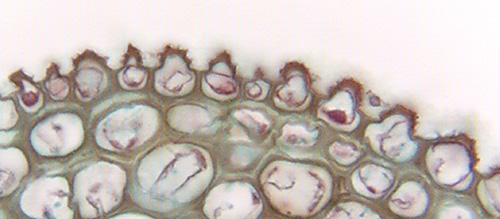
 Fig.
10.2-10. Transverse section of lilac leaf (Syringa).
These epidermis cells have an outer wall that bulges outward as a papilla, but
they also have a sculptured
cuticle. The peaks and spikes on the cuticle are so fine that they
are barely visible even at this high magnification. In a section like this, they
appear to be just spikes, but if may be that we are seeing cross sections of
ridges of cutin – we would need SEM or leaf peals to determine that. For
example, look at the SEMs of cutin ridges and projections in Figs. 10.14 and
10.16 on page 177 in Plant Anatomy (Mauseth).
Fig.
10.2-10. Transverse section of lilac leaf (Syringa).
These epidermis cells have an outer wall that bulges outward as a papilla, but
they also have a sculptured
cuticle. The peaks and spikes on the cuticle are so fine that they
are barely visible even at this high magnification. In a section like this, they
appear to be just spikes, but if may be that we are seeing cross sections of
ridges of cutin – we would need SEM or leaf peals to determine that. For
example, look at the SEMs of cutin ridges and projections in Figs. 10.14 and
10.16 on page 177 in Plant Anatomy (Mauseth).