Overview of animal viruses
Structure and replication, pathogenesis will be covered when we talk about specific diseases
Animal viruses -
Similarities to bacteriophages
Difference between animal viruses and bacteriophages:
Host cell is compartmentalized, virus must get to appropriate sites to usurp host functions
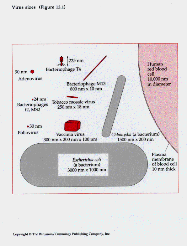
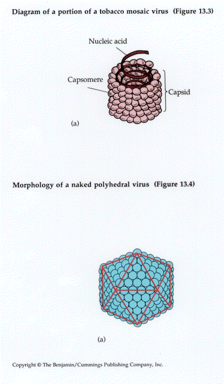
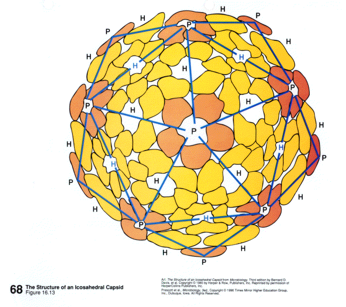
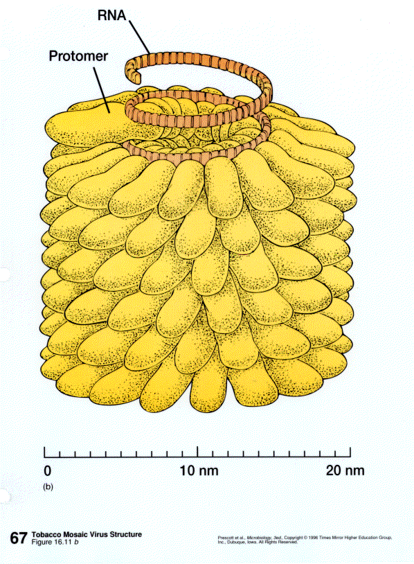
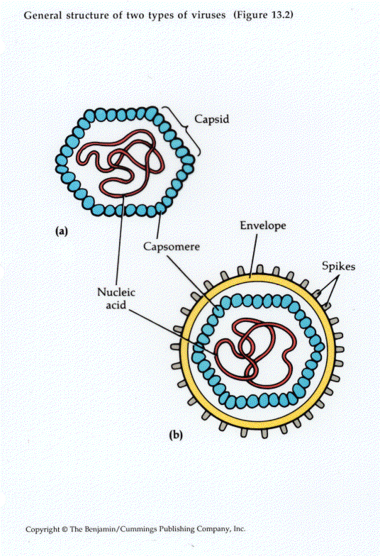
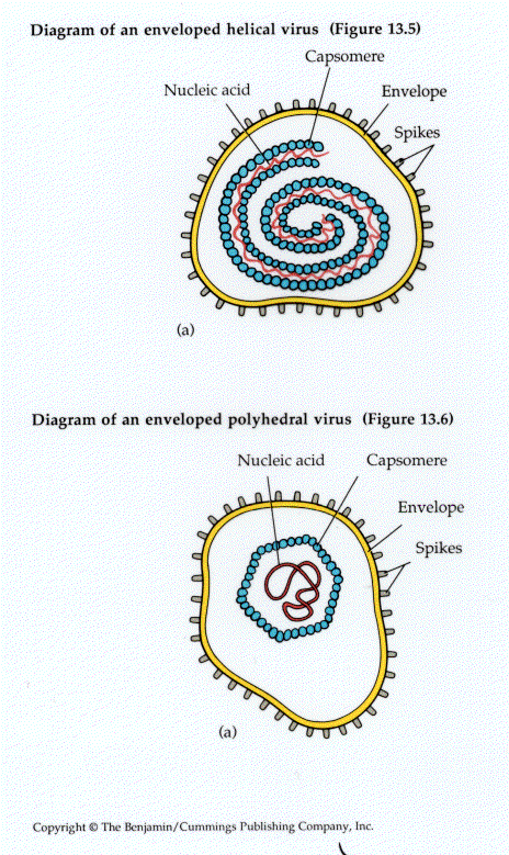
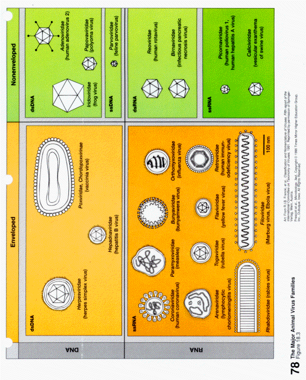
Structure of animal viruses
(Figure 3, Figure 4, Figure 5)
Viral replication
Viruses must replicate inside host cells. There is host specificity - may be species specificity or specificity for particular types of cells within the host
Steps in replication:
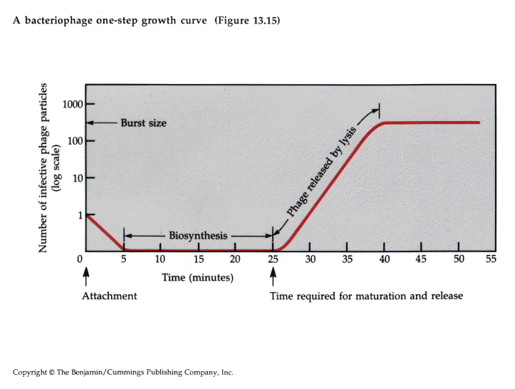
Lytic viruses show one-step growth curve
The effects of animal viruses on the host cell:
Use tissue culture to see effects of animal viruses in the laboratory
Brief overview of viral replication strategies:
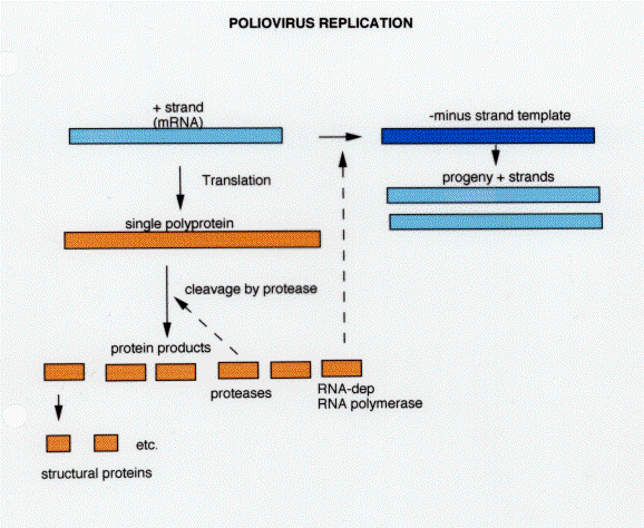
1. Positive-strand RNA virus: Figure 14
polio and hepatitis A (picornaviruses)
Positive strand means the RNA can act directly as a messenger RNA
Polio virus is a linear ssRNA, 7.5 kb, has a viral protein (VPg) covalently attached at the 5' end and a 3" poly A tail (polyA tail essential for infectivity)
RNA is translated into a single polypeptide (moncistronic) which is subsequently cleaved (posttranslational cleavage) into ~20 proteins. These include the 4 structural protein of the virus particle, VPg, the RNA dependent RNA polymerase required for replication, and a protease required for cleavage.
The RNA-dependent RNA polymerase (replicase) transcribes the viral RNA to produce the minus strand intermediate which are then transcribed to produce progeny plus strands.
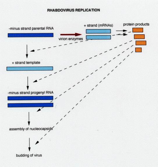
1. Negative-strand RNA virus:
rabies (rhabdovirus): Figure 15
RNA is negative strand and cannot act as mRNA, virus particles contain the RNA dependent RNA polymerase, may also have other enzymes such as RNA methylase and capping enzyme in the virion
RNA is transcribed to yield a series of mRNAs from the viral genes
RNA is also transcribed to produce a full length plus strand which serves as template for progeny negative strands.
mRNAs are translated to produce structural proteins and RNA polymerase
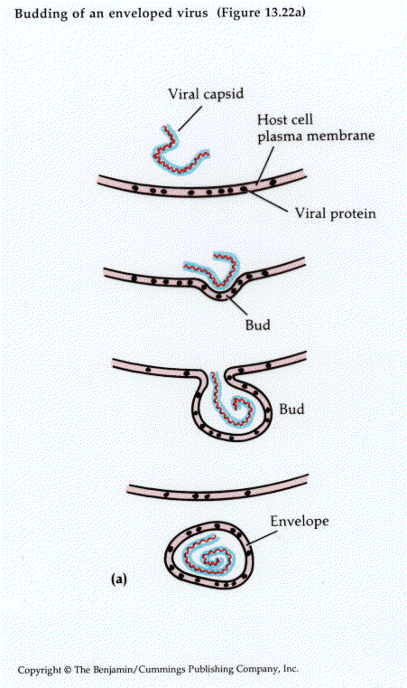
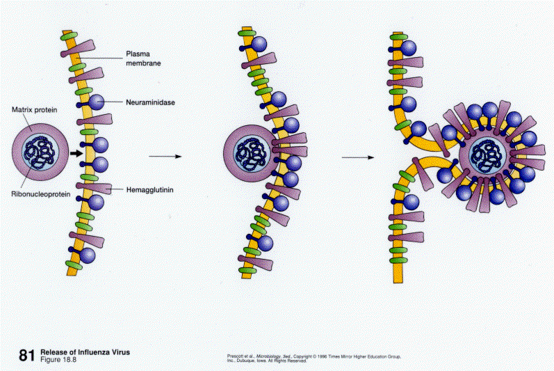
Membrane proteins are inserted into host membrane, nucleocapsids assemble and align with areas of the membrane where viral glycoproteins are inserted, virus is released by budding
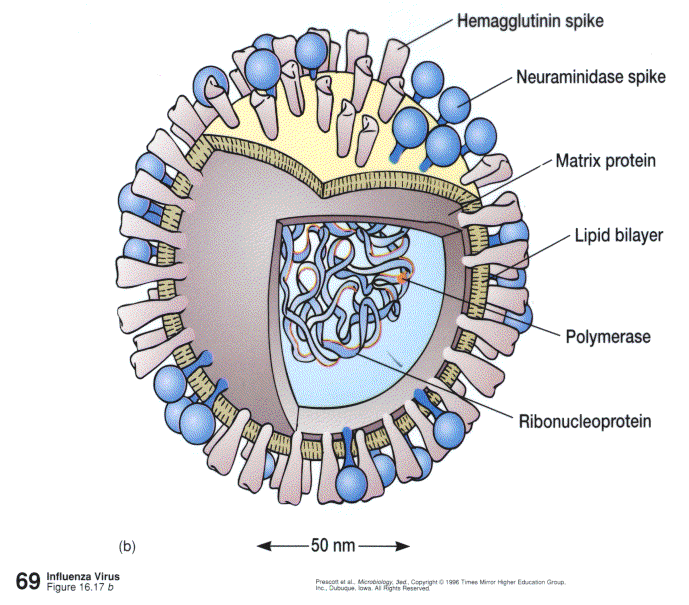
Influenza virus is also a negative stranded RNA virus. However, its genome is segmented, 8 segments encode 10 viral proteins in influenza A viruses. After entering the cell by endocytosis, the nucleocapsid migrates to the nucleus where replication occurs. Viral mRNAs are capped and polyadenylated and migrate to the cytoplasm to be translated.
Double stranded RNA viruses - Reoviruses
Double shell capsid - outer one composed of three proteins and the inner one is made up of 5 protein subunits
Particle contains RNA-dependent RNA polymerase and capping enzymes
Genome is segmented
Replication occurs in the cytoplasm, within a subviral particle. dsRNA cannot act as message so the -strand is first transcribed. mRNAs are capped. Capped +strands act as templates for progeny virus as well as message.
DNA viruses
Most of the DNA viruses are double strand (ss DNA - parvovirus)
All except the pox viruses replicate in the nucleus
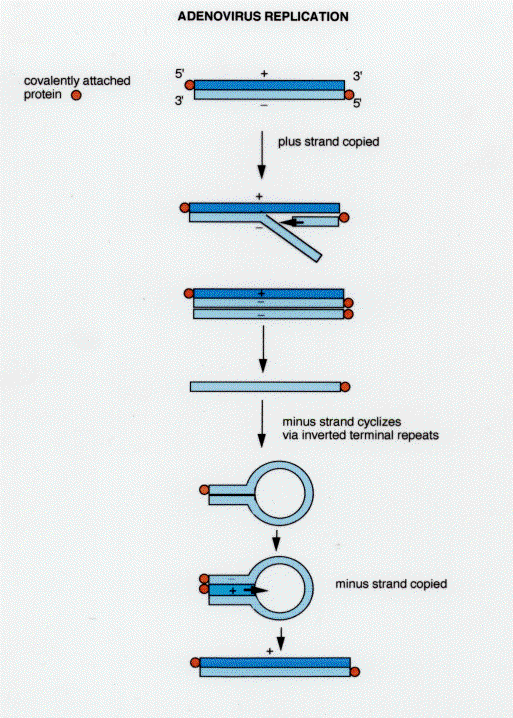
Adenovirus: Figure 16
Transparency shows relationship between activities and sites in the host cell.
dsDNA is linear and has a viral protein covalently linked to the 5" end of each strand. Replication can begin at either end but replication is asynchronous. A round of replication produces a double strand and a single strand (TRANSPARENCY). Because the ends have inverted repeats, the single strand can self anneal and be copied from the ds end.
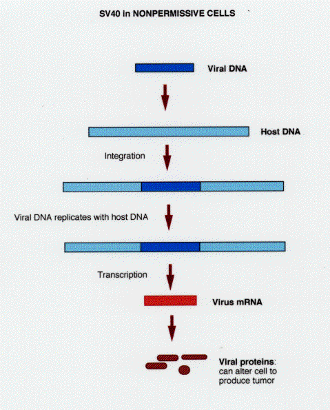
Papovaviruses (SV40) genome is a ds circular DNA: Figure 17
If the host cell is permissive < lytic infection
if host cell is nonpermissive, viral DNA may become integrated into host DNA < transformed cells
SV40 DNA replicates in the nucleus, proteins are synthesized in the cytoplasm but go back to the nucleus for assembly of the virions.
Replication proceeds in two distinct phases: early and late. Early phase results in production of T antigen which binds to the origin of replication. Late phase includes production of capsid proteins. In nonpermissive cell, the late phase is blocked and there is no replication of viral DNA. The viral DNA may be stably integrated into the host cell DNA and then replicates and can be transcribed as a cellular gene. May give rise to transformed, tumor cells.
Herpesviruses
linear dsDNA
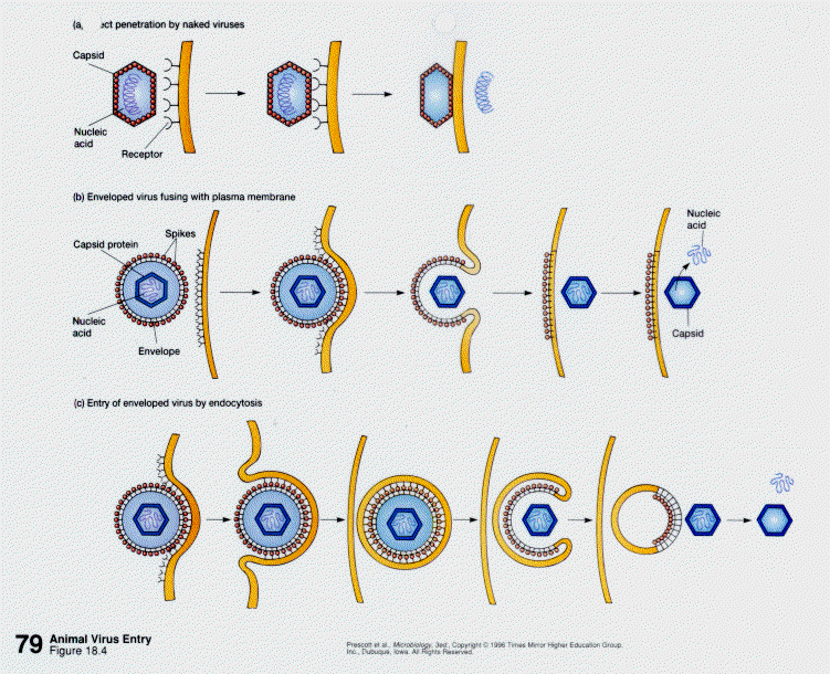
Viral envelope fuses with host cell membrane and nucleocapsid is released into the cell and transported to the nucleus. Viral DNA replication takes place in the nucleus and viral assembly also takes place here. Envelope is acquired by budding through the nuclear membrane.
Herpesviruses can remain latent in the body for long periods of time. Can be activated by stress.
Herpesviruses can be associated with cancer.
Pox viruses are very large and complex dsDNA viruses. They replicate in inclusion bodies in the cytoplasm. They encode a number of their own replication enzymes.
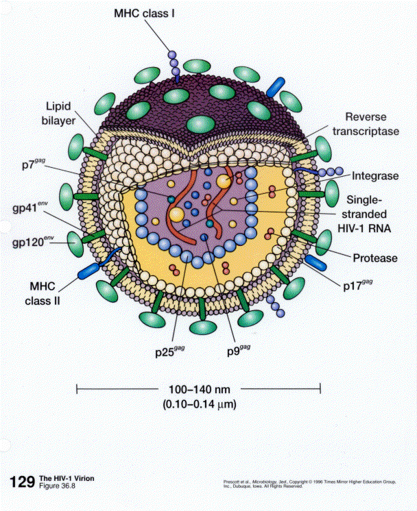
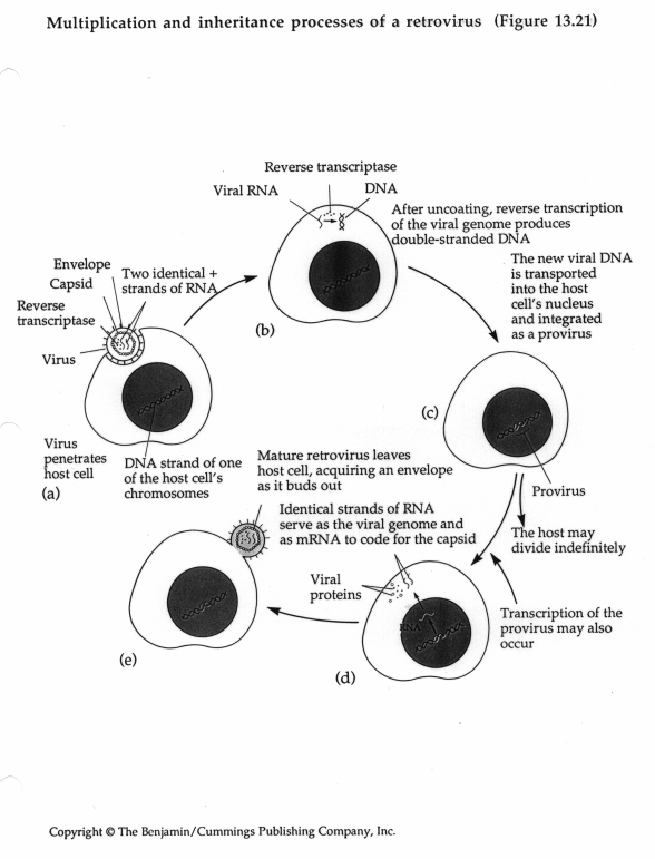
Retroviruses
Flow of information is reversed (hence the name retro) in these viruses. RNA< DNA. Use reverse transcriptase for replication. Genome consists of two identical copies of the + strand RNA held together by base pairing with tRNAs. Ends of the molecules are long terminal repeats (LTRs).
Some retroviruses carry oncogenes which can cause transformation of cells.
Major steps in replication: Figure 19
Strategies for maximizing coding capacity of small viral genomes